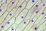
It is possible to observe the impression of leaf epidermis cells on white wood glue. The stomata and guard cells are easily visible. The regular shape of the stomata makes it an ideal specimen for practicing drawing.
Microscope info | Citizen Science | Amateur Microscopy

This category contains posts with labwork that can be done in a school.
It is possible to observe the impression of leaf epidermis cells on white wood glue. The stomata and guard cells are easily visible. The regular shape of the stomata makes it an ideal specimen for practicing drawing.

It is possible to observe the plasmolysis of cells under the microscope. When salt water is added to onion cells, then the cells will lose water due to osmosis, this can be observed.
This is one of my favorite lab activities. Onion cells are visualized using a slide projector. Using an internal reference mark, the students can calculate the actual size of onion cells. It does not require the use of microscopic equipment and can be conducted in the normal classroom (lab not required).
It is possible to enrich microorganisms such as ciliates by making a hay infusion.
Paramecia are fresh-water ciliates that make excellent microscopic specimens. They are relatively large and therefore easily observable, even under low magnification. Pond water usually does not contain sufficiently high concentrations of them. For educational purposes it is necessary to enrich them.

Crystals of organic substances make interesting microscopic specimens to be viewed under polarized light.
This is a simple preparatory technique that allows students to observe the otherwise difficult to see nucleus of onion cells. There is no need to employ, possibly harmful, DNA staining chemicals.
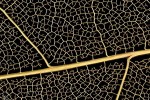
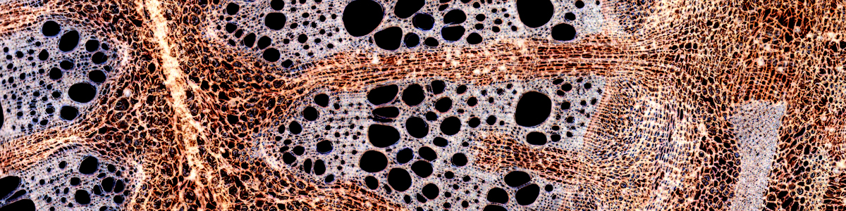
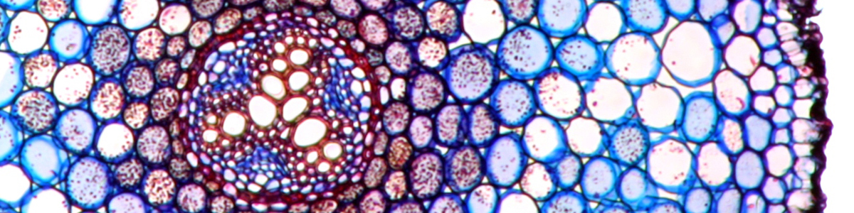
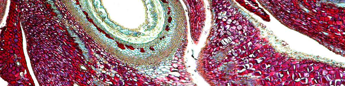
This is a simple but somewhat time-consuming preparatory technique. It is possible to isolate the vascular bundles of certain leaves and prepare them for microscopic observation. The prepared leaf veins make an ideal specimen for stereo microscopy. The microscope allows the students to perform a quality-check of their preparation.
This site uses cookies. By continuing to use the site, you agree to the use of cookies. For more information <a href= more information
The cookie settings on this website are set to "allow cookies" to give you the best browsing experience possible. If you continue to use this website without changing your cookie settings or you click "Accept" below then you are consenting to this.