Wow Jim!
A
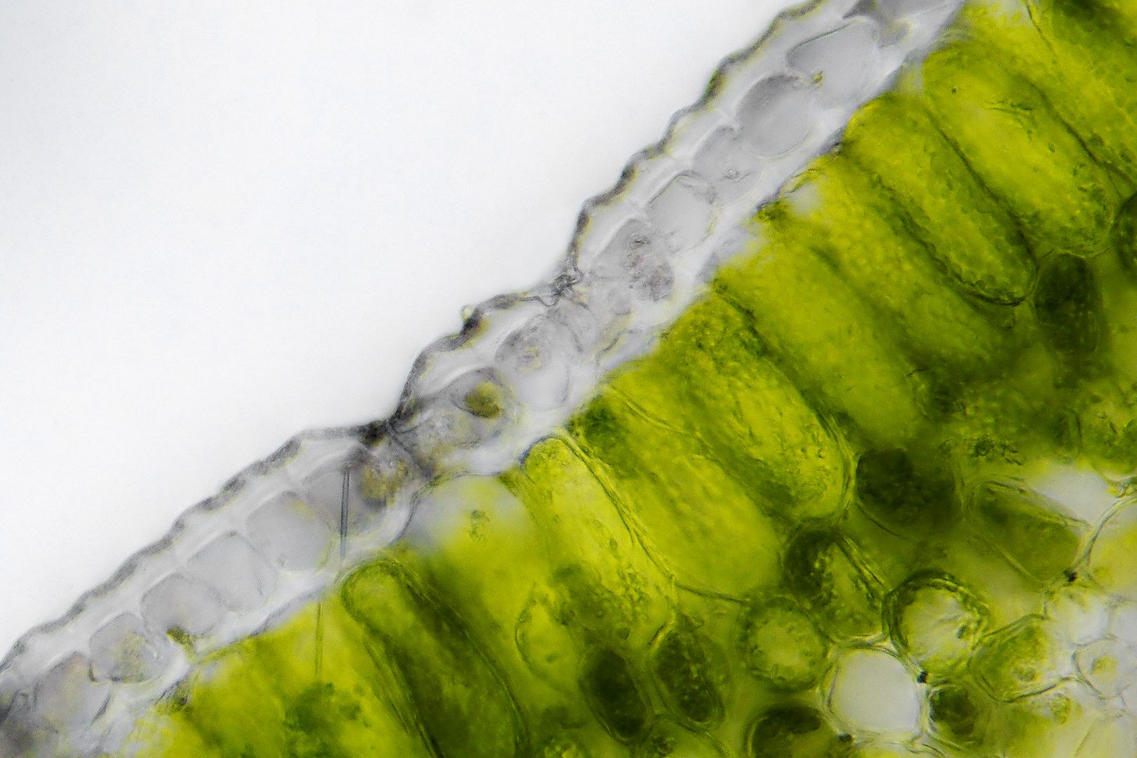
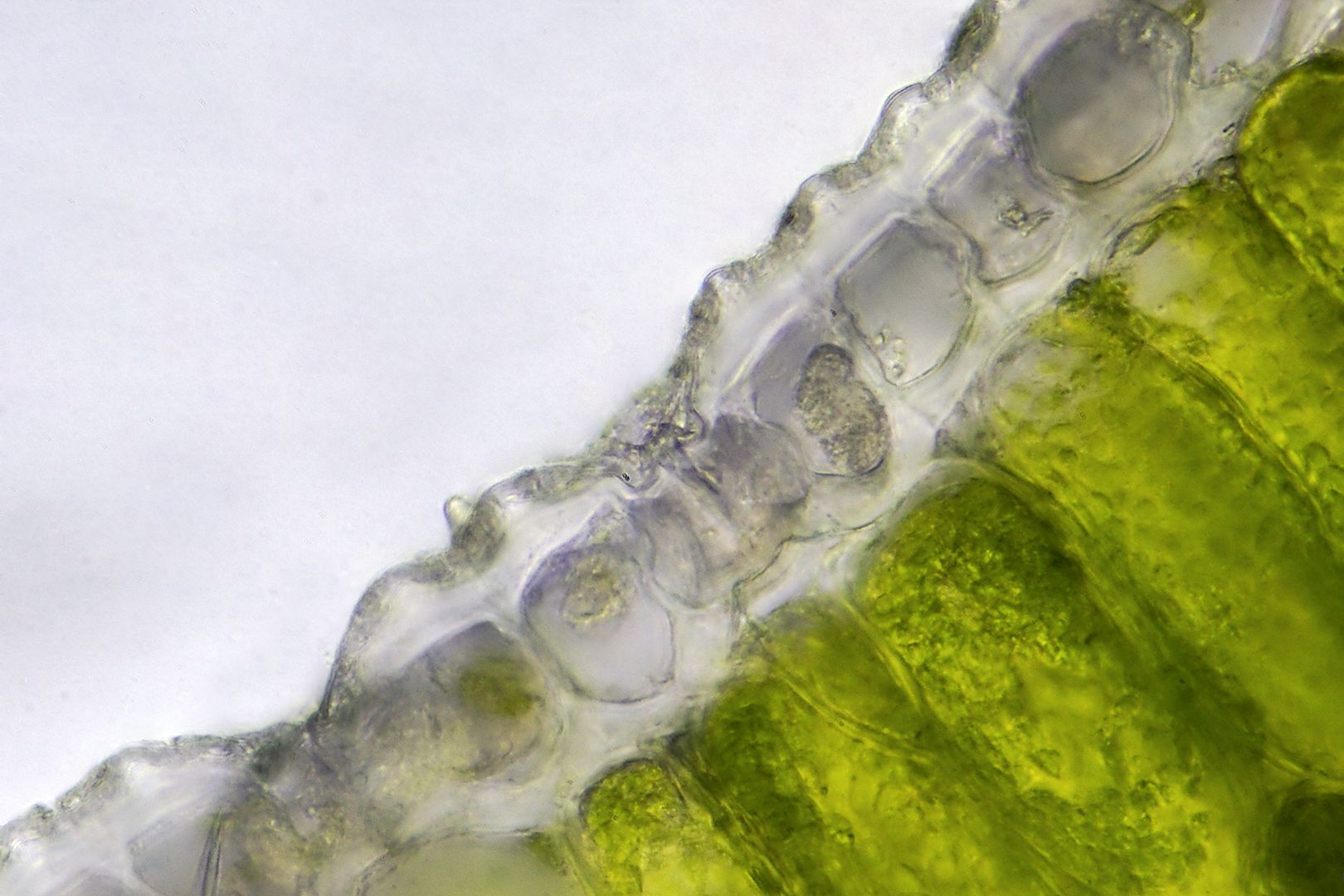
superb pair of images that really emphasize the structure as you say of the tissue. The thick cuticle is beautifully 'plump' in the live section, as is the transparent (to allow the efficient passage of light through to the chlorophyll of the palisade mesophyll below it) epidermal layer of almost cuboid cells.
The chloroplasts are clearly shown also to be lining the cells rather than 'filling' them as it were. It would be interesting to see the difference if possible between the sections of a leaf taken (very quickly to the 'scope) in bright direct sunlight and one taken on a dark/cloudy day - the very bright leaf should have it's chloroplasts in a different position to those of the leaf in dull/dark conditions....
Yes, those are definitely stomates in the epidermis too my friend.
Give us more Jim!