
Hi everyone,
I was able to enrich a population of Mayorella (correct me if my identification is wrong) and decided to experiment with it. Here I present some videos showing what happens to the amoeba when it is stained with acridine orange and irradiated with 450-490 nm light. After a few seconds of exposure the amoeba "explodes". I think this is due to the excitation of acridine orange, which then transfers part of its energy to excite oxygen to the singlet state. This reactive form of oxygen produces lipid peroxides. The peroxides lead to dramatic decomposition of the AO accumulating membranous vesicles (shown here fluorescing green), which in turn spill their contents into the cytoplasm, causing the cells to lyse suddenly.
The first frame shows a 4x accelerated video of the amoeba in DIC. The next two frames show the cellular decomposition after brief irradiation with blue light (the fluorescence videos are at 1x normal speed)
And here is another video from the same sample showing the amoeba in phase contrast (4x normal speed).
Best regards,
Wes
Exploding fluorescent amoeba
Exploding fluorescent amoeba
Zeiss Photomicroscope III BF/DF/Pol/Ph/DIC/FL/Jamin-Lebedeff
Youtube channel
Youtube channel
Re: Exploding fluorescent amoeba
Impressive and interesting experiment and demonstration !
Some questions about details:
1. Is this rapid process known from other cells or protozoans ?
2. How intense was the irradiation ? with which lamp ?
3. Would the amoeba explode if similarly irradiated but without the previous staining with AO ? or better yet, staining with a different stain that is known to be stable when exposed to the 450-490nm ?
Re: Exploding fluorescent amoeba
Thanks Hobbyist! I think the phototoxicity of acridine orange is not uncommon among protists. Initially I was studying its effects on Paramecium, however it doesn't explode but contracts and releases vesicular contents in the cytoplasm but doesn't decompose like the amoeba. I used a 100 W halogen lamp at 12 volts. I've observed the amoeba for extended periods of time in other illumination modes (phase, DIC) and nothing happens so I think the effect is mediated by AO phototoxicity.
Do you have an alternative stain for a control experiment in mind?
Do you have an alternative stain for a control experiment in mind?
Zeiss Photomicroscope III BF/DF/Pol/Ph/DIC/FL/Jamin-Lebedeff
Youtube channel
Youtube channel

Re: Exploding fluorescent amoeba
That was dramatic! Was it at normal speed?
I take it you used Interference Filters with your 100w halogen lamp. Did you take any UV precautions or just made sure to use the camera as a go between?
Additional non related question: do you normally use any IR or UV filters when using the 100w Halogen light?
Nice phase!
I take it you used Interference Filters with your 100w halogen lamp. Did you take any UV precautions or just made sure to use the camera as a go between?
Additional non related question: do you normally use any IR or UV filters when using the 100w Halogen light?
Nice phase!
Zeiss Standard WL (somewhat fashion challenged) & Wild M8
Olympus E-P2 (Micro Four Thirds Camera)
Olympus E-P2 (Micro Four Thirds Camera)
Re: Exploding fluorescent amoeba
Thanks 75'!
Yes, the fluorescence videos are at normal speed. The DIC and phase are both at 4x.
Correct, I used interference filters and a dichroic mirror attached to the fluorescence condenser with tape (had to improvise here). No UV precautions, I think the halogen bulb has a UV blocking layer, I've not formally measured any UV leaking out of it but I haven't noticed any issues (such as my retinas peeling off) following extensive hours at the microscope (its the same halogen lamp I use for normal microscopy). In fact I've been lurking around on ebay for halogen bulbs without UV protection for fluorescence microscopy in the UV range but haven't really followed up on that.
Again no anti-UV measures but I do use a KG1 IR blocking filter. The microscope has one installed at the base between the source and field diaphragm. The lamp gets incredibly hot so I think its important to protect your eyes and optics (and specimens I guess) against potentially harmful IR radiation.
Zeiss Photomicroscope III BF/DF/Pol/Ph/DIC/FL/Jamin-Lebedeff
Youtube channel
Youtube channel
Re: Exploding fluorescent amoeba
Thanks for that. Where did you source yours?Wes wrote: ↑Wed Dec 02, 2020 3:26 pm
Again no anti-UV measures but I do use a KG1 IR blocking filter. The microscope has one installed at the base between the source and field diaphragm. The lamp gets incredibly hot so I think its important to protect your eyes and optics (and specimens I guess) against potentially harmful IR radiation.
Zeiss Standard WL (somewhat fashion challenged) & Wild M8
Olympus E-P2 (Micro Four Thirds Camera)
Olympus E-P2 (Micro Four Thirds Camera)
Re: Exploding fluorescent amoeba
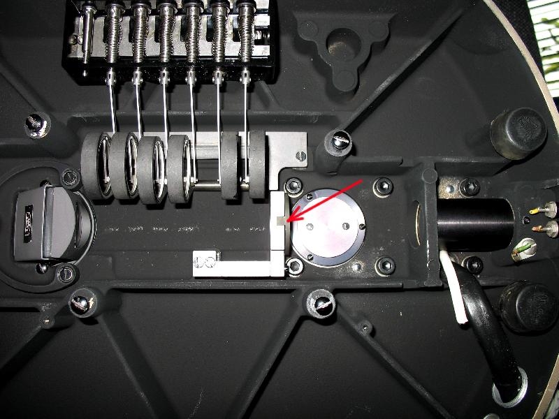
It came with the microscope, its installed by default. The photo below shows you where its placed on a Universal stand. You might be able to source one at a reasonable price on Ebay (search for KG1 heat absorbing filter).75RR wrote: ↑Wed Dec 02, 2020 4:01 pmThanks for that. Where did you source yours?Wes wrote: ↑Wed Dec 02, 2020 3:26 pm
Again no anti-UV measures but I do use a KG1 IR blocking filter. The microscope has one installed at the base between the source and field diaphragm. The lamp gets incredibly hot so I think its important to protect your eyes and optics (and specimens I guess) against potentially harmful IR radiation.
Zeiss Photomicroscope III BF/DF/Pol/Ph/DIC/FL/Jamin-Lebedeff
Youtube channel
Youtube channel
Re: Exploding fluorescent amoeba
That is interesting. Was going to put it on the field diaphragm housing filter holder ... thinking now that is probably not the best solution. Perhaps I can DIY something.
Found a seller:
https://www.galvoptics.co.uk/optical-co ... s-filters/
Zeiss Standard WL (somewhat fashion challenged) & Wild M8
Olympus E-P2 (Micro Four Thirds Camera)
Olympus E-P2 (Micro Four Thirds Camera)
Re: Exploding fluorescent amoeba
Thats a very decent price, well spotted! Keep in mind that if its too close to the source it may explode or more realistically crack from too much heat retention.75RR wrote: ↑Wed Dec 02, 2020 5:07 pmThat is interesting. Was going to put it on the field diaphragm housing filter holder ... thinking now that is probably not the best solution. Perhaps I can DIY something.
Found a seller:
https://www.galvoptics.co.uk/optical-co ... s-filters/
Zeiss Photomicroscope III BF/DF/Pol/Ph/DIC/FL/Jamin-Lebedeff
Youtube channel
Youtube channel
Re: Exploding fluorescent amoeba
I used KG3 and/or KG5 in the exit port of a 100W Hg lamp, and it indeed cracked after (I guess) around 100 hours. Indeed, the filter should be several centimeters at least from the lamp.
Re: Exploding fluorescent amoeba
Thanks for the tip. Will keep it in mind.
Zeiss Standard WL (somewhat fashion challenged) & Wild M8
Olympus E-P2 (Micro Four Thirds Camera)
Olympus E-P2 (Micro Four Thirds Camera)
-
Bruce Taylor
- Posts: 1002
- Joined: Fri Sep 04, 2015 11:34 am
Re: Exploding fluorescent amoeba
An interesting experiment.
As for the ID, my understanding is that is extremely difficult to distinguish most Mayorella species from Korotnevella, unless you have access to SEM. See the Remarks section, here: https://www.arcella.nl/genus-mayorella/ Unfortunately, if you ID to the least inclusive clade containing both organisms, the most you can say is that it's in class Discosea.
As for the ID, my understanding is that is extremely difficult to distinguish most Mayorella species from Korotnevella, unless you have access to SEM. See the Remarks section, here: https://www.arcella.nl/genus-mayorella/ Unfortunately, if you ID to the least inclusive clade containing both organisms, the most you can say is that it's in class Discosea.
Re: Exploding fluorescent amoeba
Thanks Bruce! I once asked Ferry Siemensma about an amoeba I had made a video of and he just said Mayorella.Bruce Taylor wrote: ↑Wed Dec 02, 2020 10:05 pmAs for the ID, my understanding is that is extremely difficult to distinguish most Mayorella species from Korotnevella, unless you have access to SEM.
Zeiss Photomicroscope III BF/DF/Pol/Ph/DIC/FL/Jamin-Lebedeff
Youtube channel
Youtube channel
-
Bruce Taylor
- Posts: 1002
- Joined: Fri Sep 04, 2015 11:34 am
Re: Exploding fluorescent amoeba
I would certainly accept Ferry's opinion on any amoeba you show him! When it comes to amoebae, it is my own opinion I mistrust.
In this case, there might be clues that I am unable to see & evaluate (such as the behaviour of the contractile vacuole, which apparently can be used differentiate Korotnevella from Mayorella). And of course, there are sometimes species-level characters, like the endosymbionts in Mayorella viridis, that simplify identification.
I've talked with Ferry about the growing number of amoeboid taxa that can only be identified with electron microscopy (I think heliozoans are especially problematic). He has told me that some people have actually left the hobby because of it!
There are similar issues in ciliatology, unfortunately. It's no longer possible to identify Trachelophyllum without SEM. Good old Vorticella can be quite difficult to distinguish from its cousin Pseudovorticella, without silver stain. The venerable genus Coleps has been split, and genus-level ID now requires a close look at its remarkable body-armour.