This spring I found an interesting sessile rotifer in one of my micro-aquariums, which I consider to be Collotheca trilobata (Collins 1872). I observed this animal for 19 days and during this time I filmed the original mother animal as well as the hatched juvenile animal for 4 days continuously in time lapse. This long-term observation gave me insights into the way of life of this rotifer, which I would like to share with you.
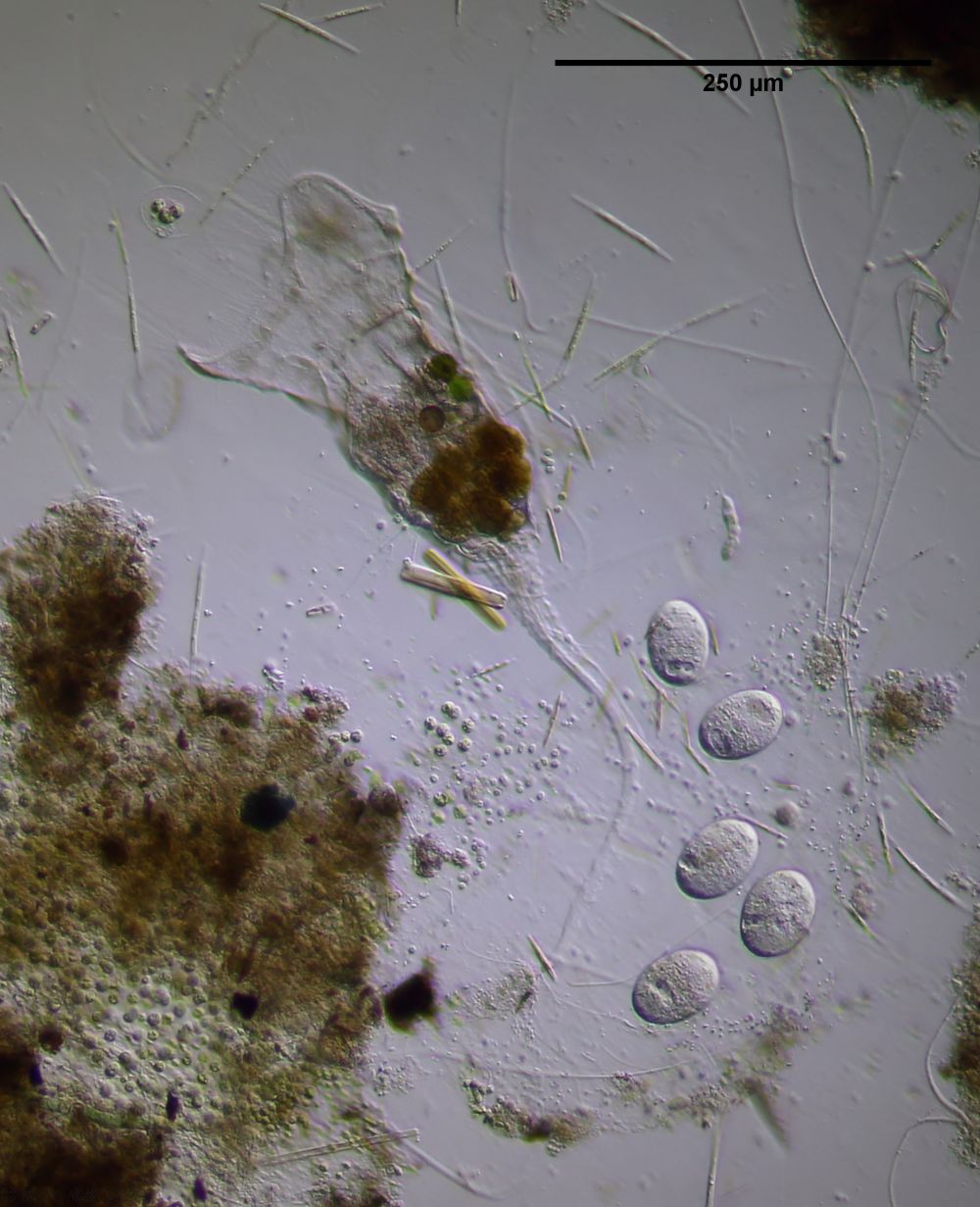
Figure 1: C. trilobata; General view of the mother with five eggs
C. trilobata is characterized by a three-lobed corona and an irregular mucous casing in which the animal lays its eggs. In order to distinguish the species from other Collotheca species with three-lobed Corona, one has to take a closer look at the Corona:
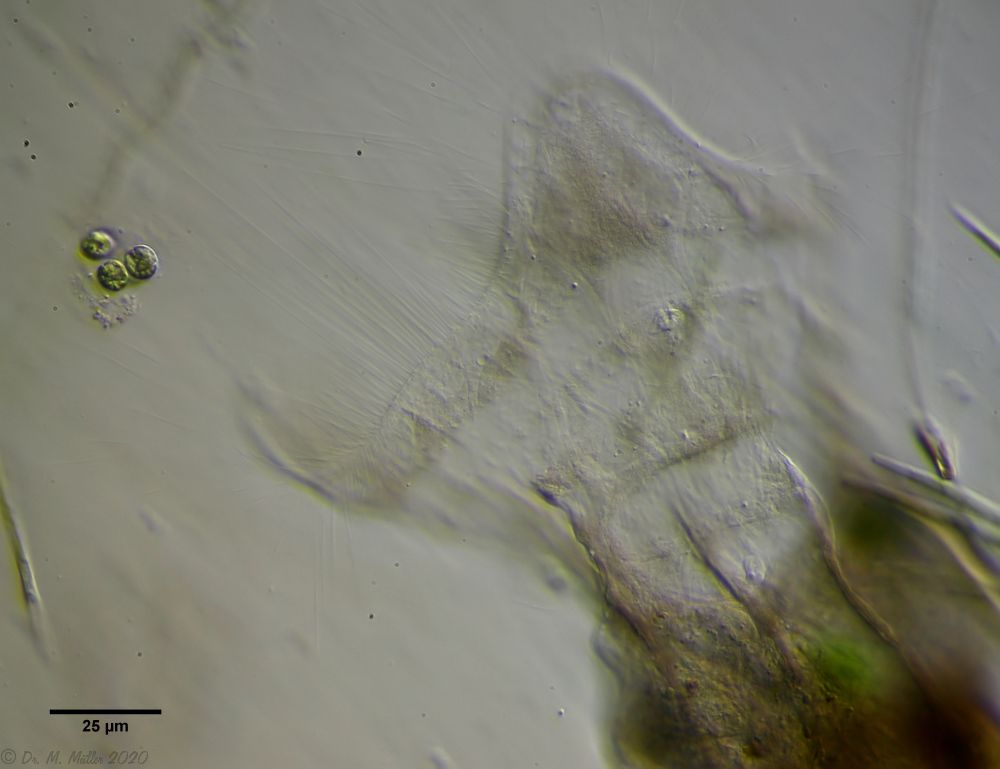
Figure 2: C. trilobata; Ciliation of the Corona
At C. trilobata the "sinusoidal" curved corona (without appendages) is surrounded by a double row of long cilia. Another typical feature is the folded foot with the short sticky handle:
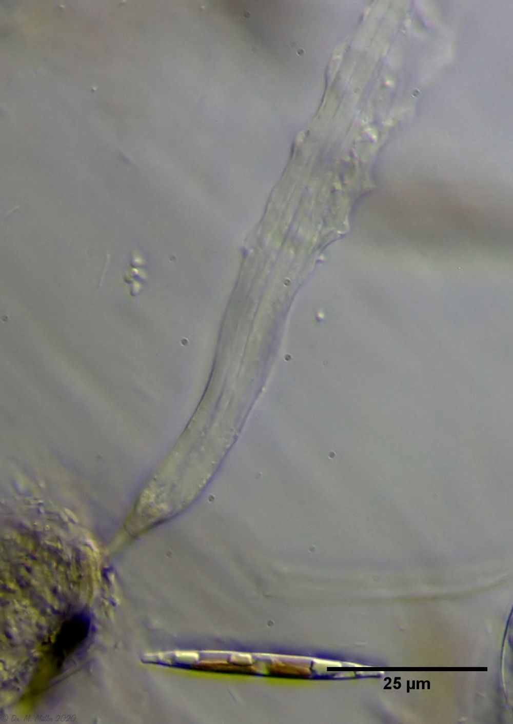
Figure 3: C. trilobata; Foot with sticky handle
Many of you will have seen rotifers of the genus Collotheca. For those who haven't had the opportunity to do so, a small film that I shot with 25x time lapse should give an impression of the "normal" behavior of these beautiful animals. I always find the rapid retraction to the housing entrance in the event of a threat particularly impressive:
Figure 4: C. trilobata; Time-lapse film on the general behavior of the animals (please click to start)
A more detailed description of these rotifers can be found in Koste (1970).
The long observation and film time enabled me to study some aspects of the behavior and development of these interesting animals more closely.
Eating behavior
The funnel-shaped crown of the animals of the genus Collotheca and its role in catching prey has often been the subject of closer investigation. It has repeatedly been speculated that the long trapping cilia on the edge of the funnel are actively used to catch prey or that they secrete attractants to attract the prey organisms. Therefore I want to discuss the feeding behavior of C. trilobata in more detail.
Let us first take a closer look at the anatomical situation of the animals' trapping and digestive systems:
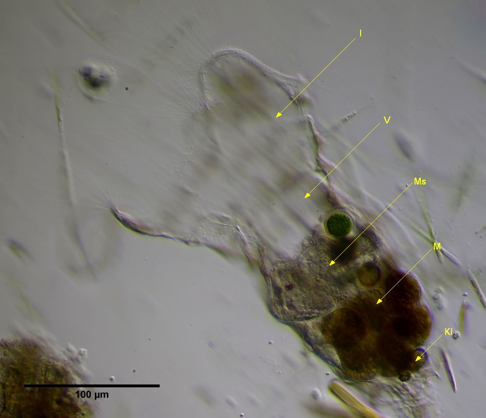
Figure 5: C. trilobata; Catching and digestive apparatus; I: interior of the funnel (infundibulum); V: vestibule; Ms: mastax sack; M: stomach; Kl: cloaca
The catching apparatus is divided into two parts, which can be separated from each other muscularly: the inner space of the catching funnel (infundibulum), which is open to the outside, is connected to the entrance of the so-called vestibulum. At the entrance there are some sensory hairs that indicate to the animal whether there is prey in the vestibule. A thin throat (pharynx) leads from the vestibule into the mastax sack, in which the animal's jaws work. The stomach, in which the actual digestion takes place, is connected to the mastax sack. The indigestible remains are collected in the cloaca as hard pellets.
In the film you can see the daughter's catch of a dinoflagellate, which I would like to discuss in advance.
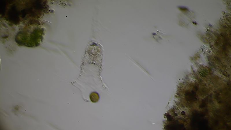
Figure 6: C. trilobata; Dinoflagellate penetrates the infundibulum
The prey enters the catching funnel on its own. The catching cilia do not actively contribute to this and only passively guide the prey towards the entrance of the funnel.
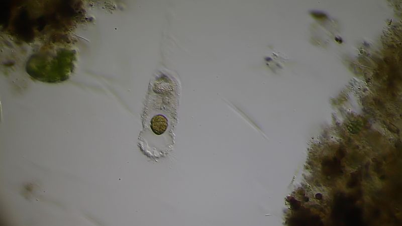
Figure 7: C. trilobata; Prey in the vestibule
The corona closes and the prey is thereby pushed further inward into the vestibule.
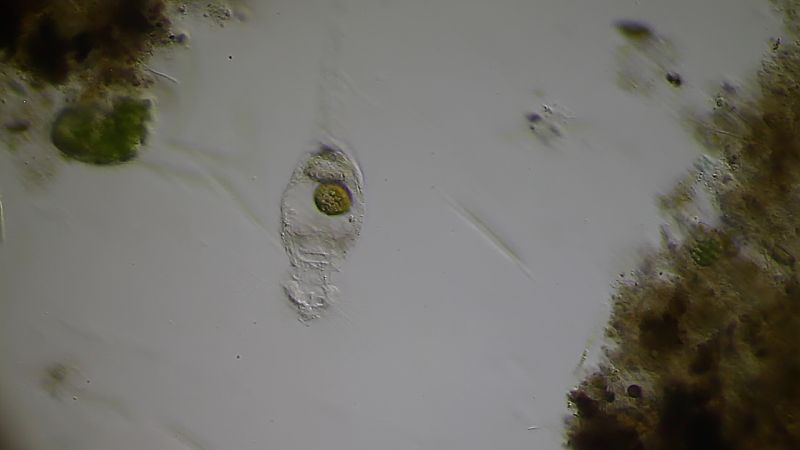
Figure 8: C. trilobata; Prey is swallowed
With a violent swallowing movement, the prey is sucked through the pharynx into the mastax sack. The mastax sack expands rapidly, sucking in the pray.
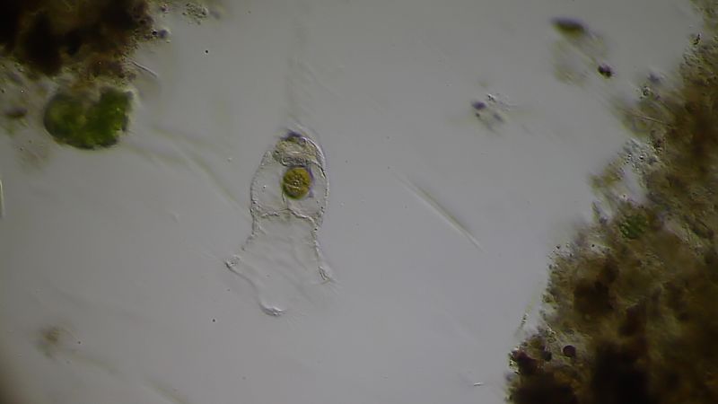
Figure 9: C. trilobata; captured prey in the mastax sack
The catching is finished and the Corona opens again for the next prey.
The prey's own movement ends within a few minutes, so I assume that the prey is already chemically killed in the mastax sack. Due to the continuous movement of the jaws and peristalsis, the prey is constantly turned over and crushed over a long period of time. In the case of the dinoflagelate shown, the comminution took longer than a day. The pulpy content of the mastax sack is then transported further into the actual stomach, where digestion takes place. The indigestible residues are first formed into loose feces pellets, which are thickened and solidified and finally added to a grain of feces in the cloaca:
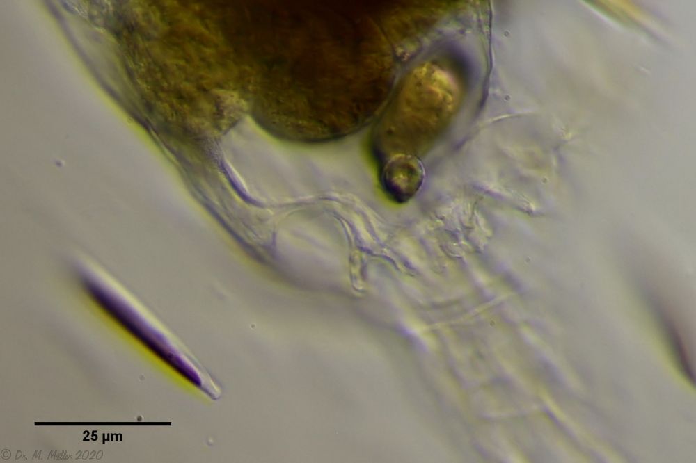
Figure 10: C. trilobata; solid fecal pellet in the cloaca
During the entire observation period, no feces were deposited, so I assume that the solid fecal pellet continues to build up. A cloaca outlet could not be observed.
I could not observe an active role of the catching cilia as described by Wright (1960) during the entire observation period. I also did not notice any evidence of a chemical attractant.
In the following film you can see some catches of prey in time lapse and in real time:
Figure 11: C. trilobata; Film on the animals' eating behavior (please click to start)
Egg development
During the observation period, the adult animal developed and laid eggs twice with an interval of approx. 12 days. I was able to film one of these eggs while it was being developed.
When I first discovered the egg, it was still flexible and relatively small. It was attached to the vitellarium and was equipped with yolk mass:
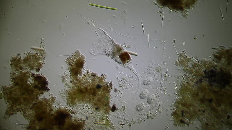
Figure 11: C. trilobata; Egg is provided with yolk mass
After a while, the still flexible and grown egg was transported into the fallopian tube:

Figure 12: C. trilobata; the egg is pushed flexibly into the fallopian tube
During its migration in the fallopian tube, the egg is rounded off and given a hard shell:

Figure 13: C. trilobata; Rounding and hardening of the egg shell
The animal withdraws to the exit of its mucous housing and the egg is deposited at the top of the housing:
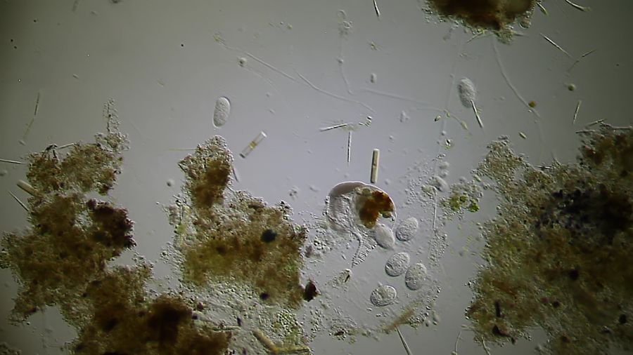
Figure 14: C. trilobata; Egg laying
Contrary to the description in Koste (1970), the egg is deposited as a single cell. I could not observe any beginning embryonic development within the mother. The corresponding cleavage divisions only took place after the egg was deposited and a polar body was ejected outside the mother's body. The egg always seems to be deposited near the exit of the case, so that the oldest egg lies at the bottom of the mucous casing.
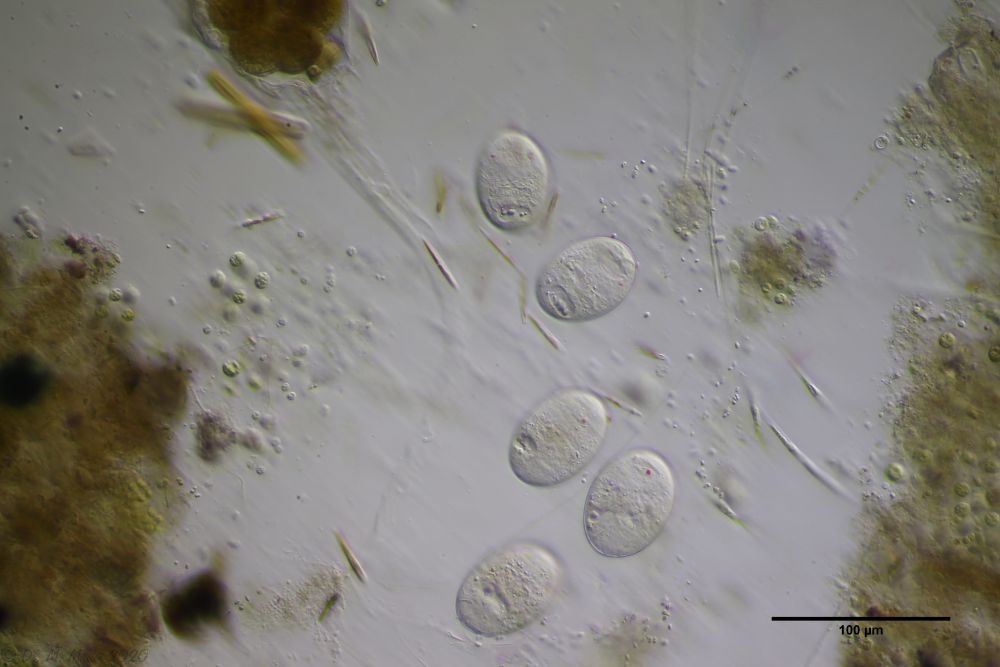
Figure 15: C. trilobata; eggs with developed embryos
The eggs develop completely but do not hatch for a long time. If the egg-laying frequency of about one egg per 12 days was maintained, the oldest egg was laid 72 days ago before I was able to observe the hatched larva.
The following time-lapse film shows egg development and the beginning of embryonic development accelerated 300 times. One minute of film corresponds to 5 hours in real time. The egg laying towards the end of the film is then shown in real time:
Figure 16: C. trilobata; Film showing the egg development of the animals
Larval development
After the embryonic development is complete, the larva remains in the egg. Eye spots and jaws can already be observed. Both movements of the larva and occasional crouching movements are visible. The "anisotropic crystalline structure" (ACS), which lie as dark granules in the cloaca of the larva, is particularly striking. This structure was interpreted by Wallace (1993) as an energy reserve for the time up to the first ingestion of food. We will later follow the fate of the ACS as the animal evolves.
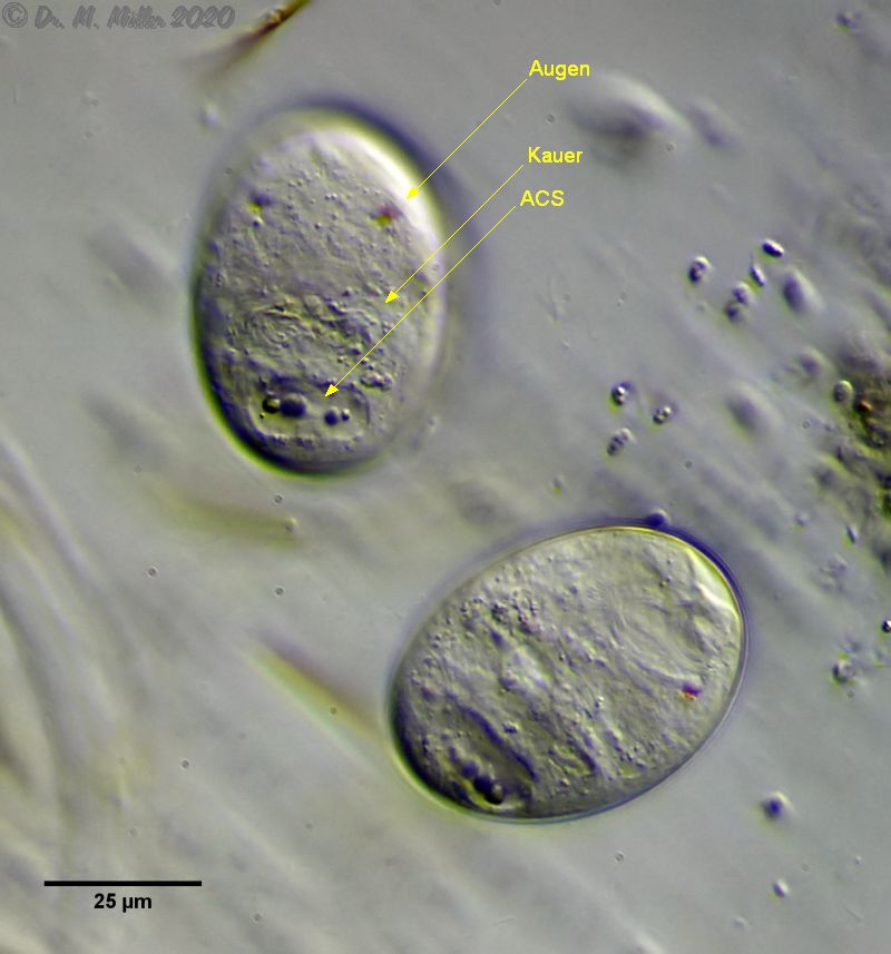
Figure 17: C. trilobata; fully developed larvae before hatching
After about 4 days of uninterrupted filming, I had actually given up hope of being able to document the larval development and only checked the state of the slide with C trilobata out of stubbornness. On the 13th day of observation, the lowest (oldest?) egg was missing in the housing:
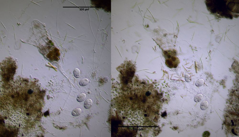
Figure 18: C. trilobata; an egg has hatched!
Whatever the trigger for the egg to hatch was, after some effort I found the larva looking for a place to settle down.
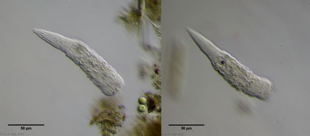
Figure 19: C. trilobata; free-swimming larva
Here, too, the ACS was easy to see. When the larva has found a place that is suitable for its later life as a sessile rotifer, it attaches itself with an adhesive thread and explores the immediate surroundings - after all, the space must offer enough space for the adult animal. I couldn't see the glue thread, but the movement of the larva clearly shows a circular path perpendicular to the animal's longitudinal axis, which I couldn't explain otherwise.
Unfortunately, I could not document the final choice of seat and could only continue with the observation a few hours later. The larva had chosen a place and settled down:
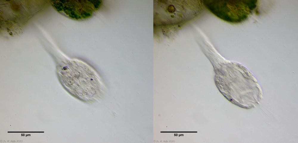
Figure 20: C. trilobata; Larva, a few hours after the settling down
From here on, the larvae development was filmed again continuously for 4 days with 25-fold time lapse.
The following film begins with the search for the free-swimming larva. After the larva has settled in place, the first eversion of the corona is shown in 25-fold time lapse. The following shows the first 50 hours of larval development in 300-fold time lapse. It is interesting to observe the ACS, which can usually be seen clearly in the film. This structure is not reduced, but grows over time. In my opinion, this structure is the beginning of the solid fecal pellet that has already been created in the egg. In any case, the ACS cannot be an "energy reserve" for the first lifetime, since it does not dissolve but grows over time!
Figure 21: C. trilobata; Film about the larval development of the animals
It was noticeable that the young animal neither produced eggs nor created a mucous casing during the 4 days of observation. Probably the housing mainly serves to protect the eggs. I never saw the adult animal retreat into the enclosure.
Best regards
Michael
Literature:
Koste W (1970): Das Rädertier-Porträt: Collotheca trilobata, ein seltenes sessiles Rädertier. Mikrokosmos 59 195
Wallace, R. L., 1993. Presence of anisotropic (birefringent) crystalline structures in embryonic and juvenile monogonont rotifers. Hydrobiologia 255/256 (Dev. Hydrobiol. 83): 71176.
Wright H G S (1960): Über den Beutefang bei Rädertieren der Gattung Collotheca. Mikrokosmos 49 228

