Trichrome staining

Here you can post pictures and videos to show others.
Post Reply
Message
Author
Naphthalene
Posts: 124
Joined: Sun Mar 29, 2015 7:34 am
Location: Moscow, Russia

Trichrome staining

#1 Post by Naphthalene » Wed Oct 21, 2015 6:05 pm

Hello everyone!
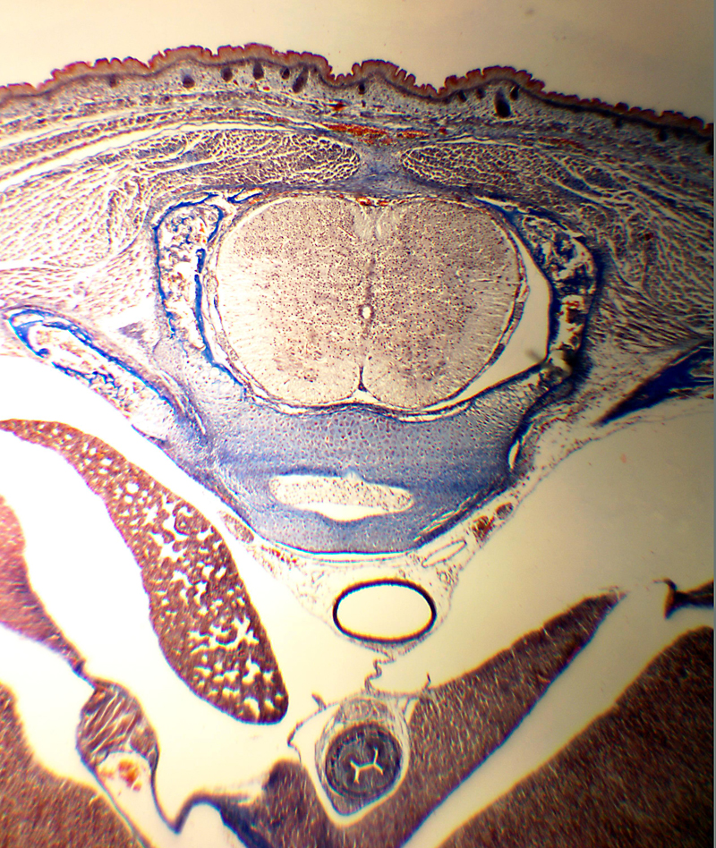
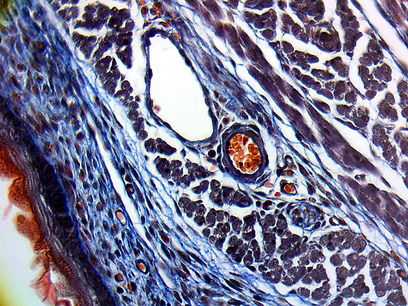
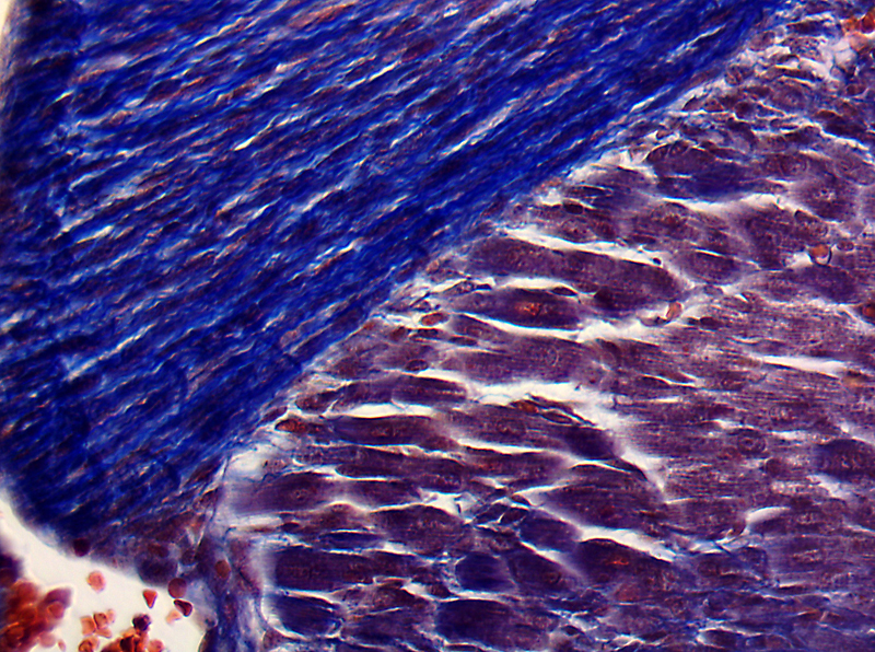
I have recently attempted to stain a mouse embryo trunk horizontal sections using Masson's trichrome method, using the IHCworld protocol with few modifications. Ponceau instead of fuchsine, some different timings. I skip the details..

Its a17 day mouse embryo, thats a secion through a belly area. You can see the spinal cord inside the future vertebrae (blue), the developing lung, liver (bottom right) and the outer skin with underlying mucle tissues. 8 micron section, Brunel rocker.

Blue is the collagen (connective tissue), red - other proteins like muscle myosin & skin keratin, dark-blue/black - cell nuclei. Erythrocytes - orange-red.

Image
Image
Image

User avatar
gekko
Posts: 4701
Joined: Sun Oct 12, 2014 7:38 am
Location: Durham, NC, USA.

Re: Trichrome staining

#2 Post by gekko » Wed Oct 21, 2015 9:49 pm

Naphthalene, excellent images with beautiful colors! I am sure a lot of expertise and hard work went into the preparation and staining, but the results speak for themselves.
Last edited by gekko on Wed Oct 21, 2015 11:29 pm, edited 2 times in total.

JimT
Posts: 3247
Joined: Fri Oct 24, 2014 1:57 pm

Re: Trichrome staining

#3 Post by JimT » Wed Oct 21, 2015 10:32 pm

Your industry continues to impress me. How big is a mouse embryo trunk horizontal section and where did you get it and how did you section it?

BTW, great images!

JimT

User avatar
mrsonchus
Posts: 4175
Joined: Tue Feb 03, 2015 9:42 pm
Location: Cumbria, UK

Re: Trichrome staining

#4 Post by mrsonchus » Wed Oct 21, 2015 11:26 pm

Beautifully clear differentiation - simply superb! :)
Very, very nice indeed. :)
John B

User avatar
Seb28
Posts: 230
Joined: Tue Nov 04, 2014 12:26 pm
Location: Ireland-Poland
Contact:

Re: Trichrome staining

#5 Post by Seb28 » Thu Oct 22, 2015 11:59 am

Very nice,I like the 2 image,very clear and sharp.
-Reichert Polyvar
-Olympus IX70
-Zeiss Photomicroscope
-Canon 600D

Naphthalene
Posts: 124
Joined: Sun Mar 29, 2015 7:34 am
Location: Moscow, Russia

Re: Trichrome staining

#6 Post by Naphthalene » Thu Oct 22, 2015 6:03 pm

Dear gekko, mrsonchus and Seb28 thank you for your kind comments!

This is a 17-18 day old mouse embryo, it is about 7 mm in diameter.

I don't work with animals myself, I get my mouse samples as a "waste product" from our animal facility, in the current example the guys were using mouse embryonic brain tissue, while the trunks were discarded. I asked them to save these trunks for me, so I have a half-dozen of them fixed in formalin in my home fridge (to scare the parents if they are to ventrue inside my "vegetable" box at the bottom :))

The paraffin embedding and sectioning were performed by standard methods. Before sectioning. the blocks were soaked in 10% ammonia for an hour, chilled with ice and immediatelly cut. Sections were stretched at 48-50C on water (<- thats the hardest part - the bone stretches ugly and gives rise to wrinkles) and mounted on gelatin coated slides.

The staining is straightforward, I simply followed the online protocol for Masson's and I did not attempt to optimize the conditions.

User avatar
Raul
Posts: 90
Joined: Tue May 12, 2015 4:55 pm
Location: Bradford, England

Re: Trichrome staining

#7 Post by Raul » Fri Oct 23, 2015 12:26 pm

Hi Naphthalene, congratulation for the samples, they are indeed with a lot of information and also they present a sweet for the eyes. I do have a question for you, did you post-process the images with a software (like photoshop or something similar) ?

Post Reply