BTW... the same gentleman that set up the web page with loads of great American Optical information also set up pages on Joseph Leidy, a scientist in the 19th century. The "entrance" to this section is here:
https://user.xmission.com/~psneeley/Personal/Leidy.htm
What is truly fascinating are the plates from his 1879 book, 'Fresh-Water Rhizopods of North America'. If you have not seen his work before you really must have a look at the pages indexed here (click on the plates for a large view). This is stunning work! (Many thanks to P.S. Neeley for making this available)
https://user.xmission.com/~psneeley/Personal/FwrPLA.htm
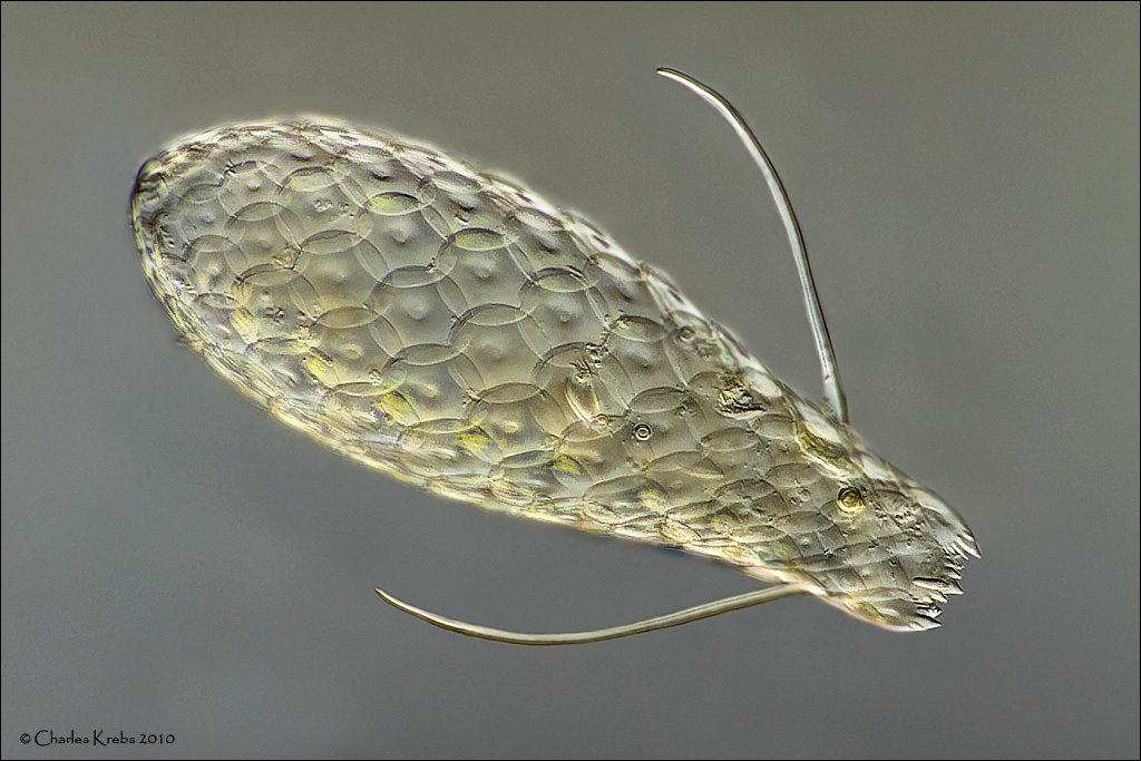
This first image is Euglypha brachiata. I collected some mosses and mud from a small stream where I have always found testate amoebae present. This is a species I really like, and I must have spent about 3 hours just looking for one and getting it onto a slide. Taken with 60X objectrive. 41 image stack. It is very transparent, so stacking was done in 4 sections to retain surface detail. It measures about 140 microns long and 50 microns wide. (0.0055 x 0.002 inch)
This second image is an Arcella (possibly A. dentata)
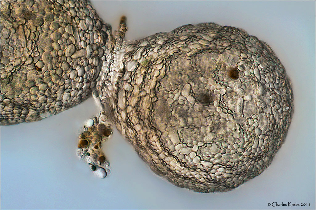
Testate amoeba usually reproduce via asexual binary fission. In the process, a "daughter" test is produced around a cytoplasmic bud that exits the original test during the process. This image is near the completion of a fission of Netzelia tuberculata.
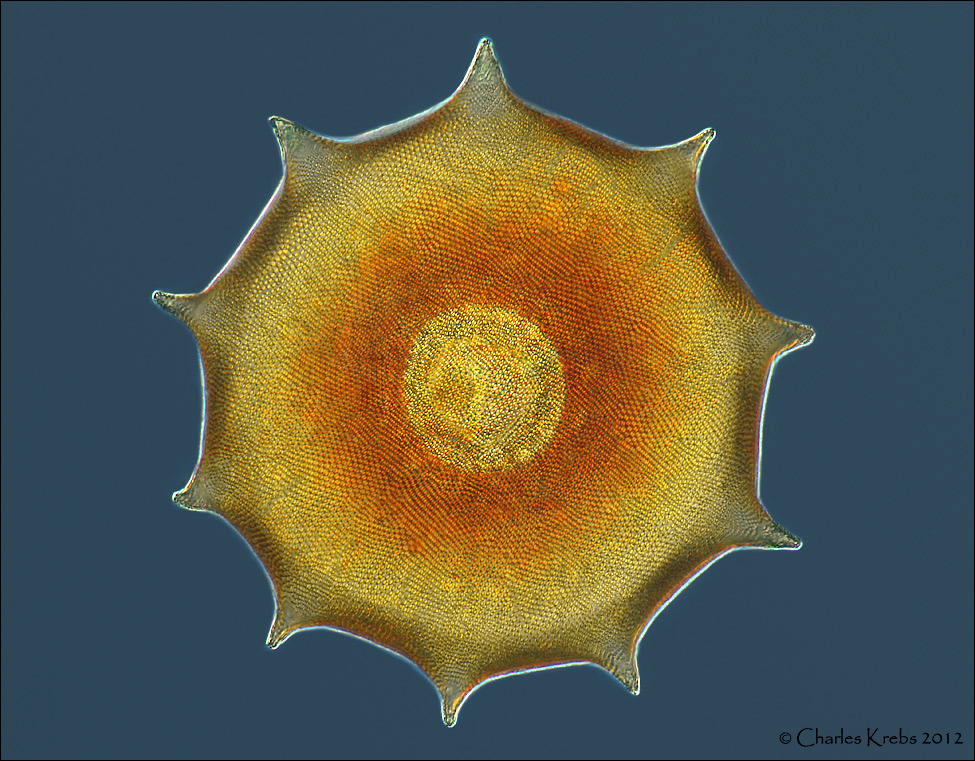
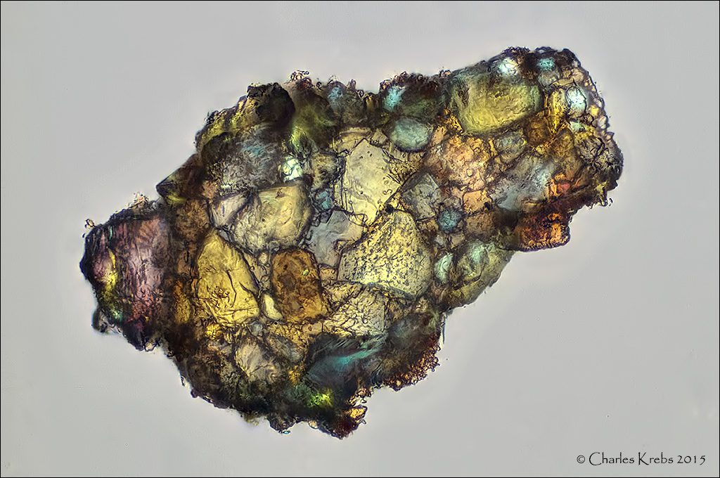
Many testate amoeba make their tests from "found" materials such as tiny grains of sand. For fun I wanted to see what one would look like under cross-polarized light.
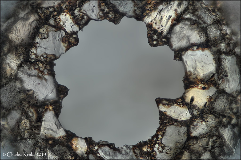
This last image is looking directly into the opening aperture of a Difflugia sp. test.