Testate Amoeba
Posted: Sat Aug 13, 2016 8:52 pm
Kurt's recent foraminifera shots reminded me how fascinated I was with amoeboid protists that made "shells" or "tests". For a very long time an amoeba was, to me, a little blob of protoplasm that oozed about. When I began finding testate amoeba I was really amazed. Hard for me to fathom such wonderful little "containers" built by such a lowly creature. I spent a lot of time searching for them. Below are some images.
BTW... the same gentleman that set up the web page with loads of great American Optical information also set up pages on Joseph Leidy, a scientist in the 19th century. The "entrance" to this section is here:
https://user.xmission.com/~psneeley/Personal/Leidy.htm
What is truly fascinating are the plates from his 1879 book, 'Fresh-Water Rhizopods of North America'. If you have not seen his work before you really must have a look at the pages indexed here (click on the plates for a large view). This is stunning work! (Many thanks to P.S. Neeley for making this available)
https://user.xmission.com/~psneeley/Personal/FwrPLA.htm
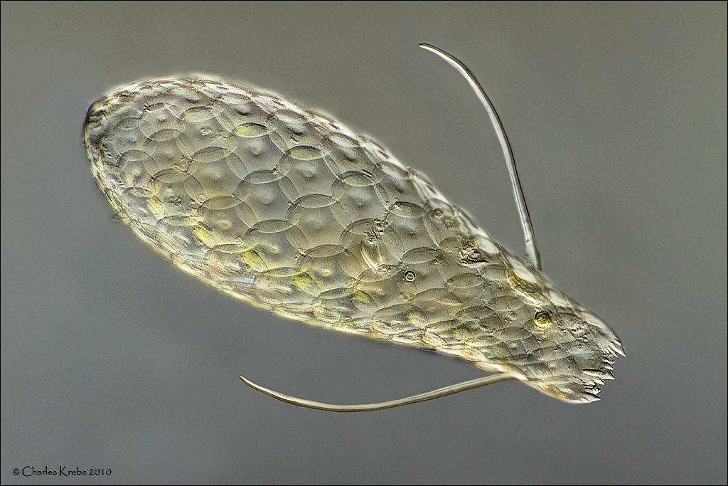
This first image is Euglypha brachiata. I collected some mosses and mud from a small stream where I have always found testate amoebae present. This is a species I really like, and I must have spent about 3 hours just looking for one and getting it onto a slide. Taken with 60X objectrive. 41 image stack. It is very transparent, so stacking was done in 4 sections to retain surface detail. It measures about 140 microns long and 50 microns wide. (0.0055 x 0.002 inch)
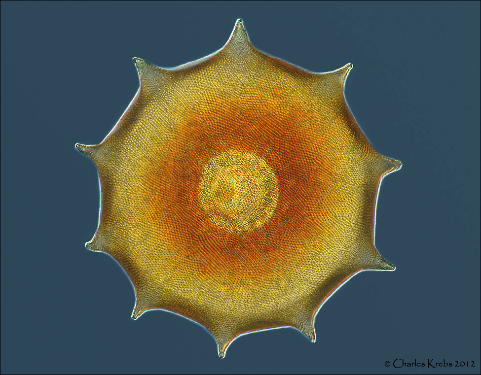
This second image is an Arcella (possibly A. dentata)
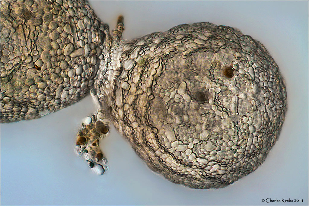
Testate amoeba usually reproduce via asexual binary fission. In the process, a "daughter" test is produced around a cytoplasmic bud that exits the original test during the process. This image is near the completion of a fission of Netzelia tuberculata.
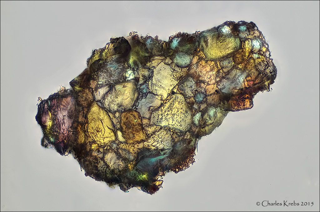
Many testate amoeba make their tests from "found" materials such as tiny grains of sand. For fun I wanted to see what one would look like under cross-polarized light.
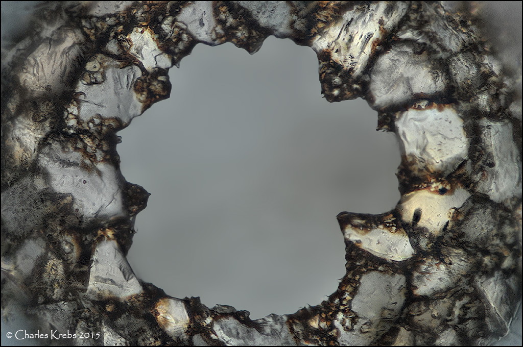
This last image is looking directly into the opening aperture of a Difflugia sp. test.
BTW... the same gentleman that set up the web page with loads of great American Optical information also set up pages on Joseph Leidy, a scientist in the 19th century. The "entrance" to this section is here:
https://user.xmission.com/~psneeley/Personal/Leidy.htm
What is truly fascinating are the plates from his 1879 book, 'Fresh-Water Rhizopods of North America'. If you have not seen his work before you really must have a look at the pages indexed here (click on the plates for a large view). This is stunning work! (Many thanks to P.S. Neeley for making this available)
https://user.xmission.com/~psneeley/Personal/FwrPLA.htm
This first image is Euglypha brachiata. I collected some mosses and mud from a small stream where I have always found testate amoebae present. This is a species I really like, and I must have spent about 3 hours just looking for one and getting it onto a slide. Taken with 60X objectrive. 41 image stack. It is very transparent, so stacking was done in 4 sections to retain surface detail. It measures about 140 microns long and 50 microns wide. (0.0055 x 0.002 inch)
This second image is an Arcella (possibly A. dentata)
Testate amoeba usually reproduce via asexual binary fission. In the process, a "daughter" test is produced around a cytoplasmic bud that exits the original test during the process. This image is near the completion of a fission of Netzelia tuberculata.
Many testate amoeba make their tests from "found" materials such as tiny grains of sand. For fun I wanted to see what one would look like under cross-polarized light.
This last image is looking directly into the opening aperture of a Difflugia sp. test.