
I just received some cleaned Diatoms in a small tube that I ordered from the Netherlands for 10 euros per tube. I ordered them from their web site:
https://www.diatoms.nl/search.html?sear ... 4178ee6bd9
I ordered both samples that they offered from Ugchelen and Renkum - they come in 2 ml tubes and I received them in about 3 weeks which is not bad considering I am located in Calgary, AB Canada.
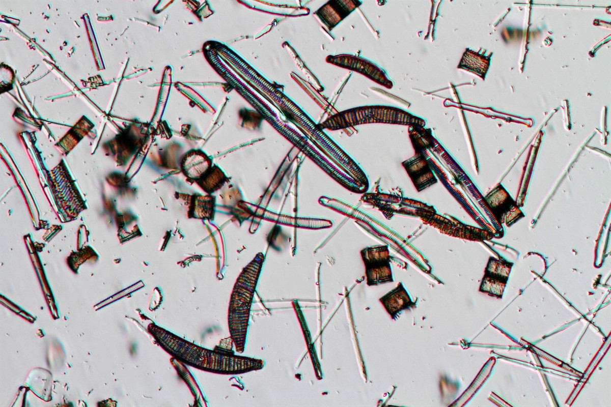
I immediately diluted some sample in water and put them on slides and took a few photos and I am quite excited about the results. I will be making some strew slides then I will try some simple arrangements. Prepared Diatoms slides are not cheap and having RAW diatoms is an alternative. Of course one can collect Diatoms locally and clean them with bleach, acid and hydrogen peroxide which I plan to do in the spring, but this is a good way to get introduced to Freshwater Diatoms and at a reasonable cost and learn to make your own slides.
For high res photos I used Photoshop to focus stack about 5-10 photos, I recorded the images using a Nikon D500 and a Zeiss Axioscope microscope and free Digicam control software to capture the images on my laptop computer. The highest resolution photo was taken with a 40X objective the central raphe or line is 0.9 microns wide.
Diatoms are one of the most beautiful subjects one can study with a microscope and I never get tired of looking at them. I am waiting on some RAW Radiolarian samples.
Diatom Samples from the Netherlands.
Diatom Samples from the Netherlands.
- Attachments
-
- 22focusstack_withbar1024.jpg (133.45 KiB) Viewed 6458 times
-
- 19Focusstack1024.jpg (64.16 KiB) Viewed 6458 times
-
- Strew DIC microscopy
- 36focusstackstrew1024.jpg (103.05 KiB) Viewed 6458 times
-
- Strew in Bright field microscopy about 50X
- BFstrew10Xobj.1024jpg.jpg (112.68 KiB) Viewed 6458 times

Re: Diatom Samples from the Netherlands.
I got a sample from them labeled Renkum Netherlands that looks very much like these.
Radazz
Radazz
Arnold, Missouri
Olympus IX70
Olympus BX40
Olympus SZ40
Olympus IX70
Olympus BX40
Olympus SZ40

Re: Diatom Samples from the Netherlands.
Very nice images!
That is a good idea getting cleaned diatoms to practice with.
Do you have a manipulator yet? Can't help wondering if you managed to get all of the diatoms off your wet slide ...
By the way the central raphe measurement you mention is a little optimistic. Although the math undoubtedly gives this number max resolution in light microscopy is around 2µm for a dry objective.
This is from the Measuring with a microscope page at Rice University: http://www.ruf.rice.edu/~bioslabs/metho ... uring.html
Last edited by 75RR on Fri Jan 04, 2019 8:10 am, edited 1 time in total.
Zeiss Standard WL (somewhat fashion challenged) & Wild M8
Olympus E-P2 (Micro Four Thirds Camera)
Olympus E-P2 (Micro Four Thirds Camera)
-
redflanker

- Posts: 47
- Joined: Mon Nov 12, 2018 9:48 am
- Location: Kunming,China
Re: Diatom Samples from the Netherlands.
Is it in sea water or fresh water?What water do you dilute the sample with?
Microscope:Zeiss Primo star with phase contrast
Camera:Cannon 90D
Amateur user
Camera:Cannon 90D
Amateur user
Re: Diatom Samples from the Netherlands.
I wonder why the page from Rice University says so. Formula for microscope resolution yield values of around 0.5 micrometer or better for NAs of 0.65-0.75, which are still 40X dry objectives (not to mention Planapo 40X0.95, but the University probably refers to student microscopes and lower NA values of the 40X objectives). For green light that is. Perhaps the Rice University lab experience is that the microscopes are not well aligned. Or that the contrast is too poor and affects the observed resolution.75RR wrote:By the way the central raphe measurement you mention is a little optimistic. Although the math undoubtedly gives this number max resolution in light microscopy is around 2µm for a dry objective.
This is from the Measuring with a microscope page at Rice University: http://www.ruf.rice.edu/~bioslabs/metho ... uring.html
Re: Diatom Samples from the Netherlands.
I think the difference is between theoretical and practical resolution as achieved by students in a University Lab with white light.Hobbyst46 wrote:I wonder why the page from Rice University says so. Formula for microscope resolution yield values of around 0.5 micrometer or better for NAs of 0.65-0.75, which are still 40X dry objectives (not to mention Planapo 40X0.95, but the University probably refers to student microscopes and lower NA values of the 40X objectives). For green light that is. Perhaps the Rice University lab experience is that the microscopes are not well aligned.75RR wrote:By the way the central raphe measurement you mention is a little optimistic. Although the math undoubtedly gives this number max resolution in light microscopy is around 2µm for a dry objective.
This is from the Measuring with a microscope page at Rice University: http://www.ruf.rice.edu/~bioslabs/metho ... uring.html
Zeiss Standard WL (somewhat fashion challenged) & Wild M8
Olympus E-P2 (Micro Four Thirds Camera)
Olympus E-P2 (Micro Four Thirds Camera)
Re: Diatom Samples from the Netherlands.
Hi - to answer your questions - I am confident of the resolution of the raphe being 0.9 microns with 40X dry objective. I determine the magnification on the digital image using a micrometer slide and eyepiece ocular to measure the length of the Diatom and and then I use Photoshop to measure the number of pixels across the rahe and convert it to size and I believe its accurate. The 40X objective resolution is = wavenlength\2NA according to Abbe. There is a short article here showing resolution is about 0.42 microns with green light so my measurement is realistic.
http://www.microscopy-uk.org.uk/mag/art ... tion-3.pdf
I posted an article on my web site showing Diatom photos by Redmayne in 1877 and he shows incredible resolution with dry objectives even back then and captured the images on gelatin prints. https://www.canadiannaturephotographer. ... atoms.html
When diluting the diatom samples I just used a bit of distilled water. I am going to make some permanent slides using Naphrax and also Canada Balsam today. I know that using a higher refractive index should increase the overall contrast, though the contrast wasn't bad in water. DIC seems to really help accentuate the contrast.
Yes I have a used Prior micromanipulator which I purchased used from Ebay, and I will make some microneedles from glass tubing and try manipulating the Diatoms. I have done some micromanipulation work before with live cells. My biggest concern is getting the diatoms to stick to the needle and then release them and have them stick to the coverslip. Steve Beats has a great article with lots of tips on Diatom arranging - see download: https://www.microscopy-uk.org.uk/mag/ar ... anging.pdf
Also if interested see: https://sciphotoclass.cias.rit.edu/word ... or-Web.pdf - both articles contains some useful information about Diatom arranging.
I have done Diatom arranging by extracting and copying images using Photoshop which is easy, but I would like to try the real thing even though I know it will tax my patience and probably take me months to get results.
Thank you for your comments
-
ImperatorRex

- Posts: 571
- Joined: Fri Mar 30, 2018 4:12 pm
- Location: Germany
- Contact:
Re: Diatom Samples from the Netherlands.
Hi Rob,
great images. How did you setup the DIC color contrast. By using a Lamda plate?
great images. How did you setup the DIC color contrast. By using a Lamda plate?
Re: Diatom Samples from the Netherlands.
Hi Jochen - I used a full 550 nm wave plate (full lambda plate) and when I turn the Zeiss DIC prisms above the objective I can achieve different coloured backgrounds with DIC. The wave plate or a quartz wedge also works with a polarizing microscope as you probably already know. I have found that mica slices and even scotch tape can mimic wave plates and produce coloured backgrounds when placed between polarizers - but I am getting off topic. I can convert the DIC scope to a polarizing microscope by removing the DIC prisms adding a polarizer above the light source and setting the condenser to bright field.
I have been trying to enhance low birefringence specimens like Diatoms using a polarized microscopy technique developed by Dr. Michael Shribak called Polychromatic polarization - but his science paper and his patent leave out important details like what type of quarter wave plate he uses (achromatic?) or z crystals (type and source) and how does his polarization state generator work to generate multiple colours. I have tried all combinations of polarizers and wave plates without any luck.
He is able to get multicolours from Diatoms viewed in polarized light. I emailed him and hoping he will clarify. If you have not seen this technique check out this paper: https://www.nature.com/articles/srep17340 if interested.
DIC is expensive and I had to wait a long time to purchase it. I think in the future we will be able to simulate DIC, Phase contrast and other techniques using software - something I have been following - this will bring these techniques into the hands of amateurs and a much larger audience.
Cheers
Rob
Re: Diatom Samples from the Netherlands.
Potentially very interesting, glass is not birefringent and diatoms fairly resemble glass.RobBerdan wrote: have been trying to enhance low birefringence specimens like Diatoms...
About arranging diatoms: here is a link to a most comprehensive live description, "Doing diatoms a different way" by forum member Charles:
viewtopic.php?t=4227
Even a video clip which demonstrates the effort and patience that are neccessary...
An article by Webb from 2015 (I think) describes condenserless phase contrast and other contrast methods by means of LED rings; but I do not know how easy it is for amateurs to make and apply it. There are also microscope developments that rely on tiny holes instead of objectives...I think in the future we will be able to simulate DIC, Phase contrast and other techniques using software - something I have been following - this will bring these techniques into the hands of amateurs and a much larger audience.
Thanks for posting the beautiful black and white photographs of diatoms from 1877!
-
ImperatorRex
- Posts: 571
- Joined: Fri Mar 30, 2018 4:12 pm
- Location: Germany
- Contact:
Re: Diatom Samples from the Netherlands.
Thanks Rob for the details.
The Polychromatic polarization article is really interesting, I did not focus much on the theory but had a quick look on the sample images provided. Results look quite psychodelic and it probabely needs some time and experience to understand how to interprete the different colors.
DIC has become much more feasible then before - respectively if you look at the older scope for finite optics. So I have seen recently a complete DIC set for less then 500 USD on ebay.

Re: Diatom Samples from the Netherlands.
Rob,
Michael Shribak has more than one patent:
http://www.mbl.edu/bell/current-faculty/shribak-lab/
Maybe the embodiments (details) you were looking for are in his previous patent(s), rather than the one you read?
A patent examiner usually won't allow a patent to be issued, if it lacks details significantly; but then some patents do not contain enough details for a hobbyist at home (even a PhD like you) to repeat them exactly. Also too much details = limited protection scope (not so nice for inventors), in general.
I read that Nature paper, but could not follow his optical theory/math (which I am poor at). He must has something really novel to be able to publish in Nature though.

Re: Diatom Samples from the Netherlands.
Hei if you're still interested in this manipulation, we've recently sorted out the gory details of Shribak's method in another thread, and found out that it can also be made by stacking with a normal Polarizing scope; no extra equipment! Ok for diatoms if they don't swim away while we are stacking.RobBerdan wrote: ↑Fri Jan 04, 2019 8:36 pmI have been trying to enhance low birefringence specimens like Diatoms using a polarized microscopy technique developed by Dr. Michael Shribak called Polychromatic polarization - but his science paper and his patent leave out important details like what type of quarter wave plate he uses (achromatic?) or z crystals (type and source) and how does his polarization state generator work to generate multiple colours.
https://www.microbehunter.com/microsco ... t=13077
short cut, post 17