
Planapo 63x/1.4, DIC, 25µm diameter, marine sample, 17 image stack in Photoshop
Cylindrical diatom attached to an alga that is coated with small oval diatoms
Cylindrical Diatom
Cylindrical Diatom
- Attachments
-
- Cylindrical Diatom--.jpg (306.39 KiB) Viewed 4748 times
Zeiss Standard WL (somewhat fashion challenged) & Wild M8
Olympus E-P2 (Micro Four Thirds Camera)
Olympus E-P2 (Micro Four Thirds Camera)
Re: Cylindrical Diatom
Here is what looks to be a spherical one from the same session.
Planapo 63x/1.4, DIC, 30µm diameter, Marine sample, 15 image stack in Photoshop.
Planapo 63x/1.4, DIC, 30µm diameter, Marine sample, 15 image stack in Photoshop.
- Attachments
-
- Spherical Diatom.jpg (154.37 KiB) Viewed 4715 times
Zeiss Standard WL (somewhat fashion challenged) & Wild M8
Olympus E-P2 (Micro Four Thirds Camera)
Olympus E-P2 (Micro Four Thirds Camera)
-
gconcepcion
- Posts: 14
- Joined: Mon Jan 07, 2019 11:39 pm
Re: Cylindrical Diatom
Great images! To be honest, the second photo of the spherical protist looks like a dinoflagellate to me. Maybe in the genus Alexandrium, or Protoceratium? Or a Gymnodinoid? Would need to see more individuals and a view of the sulcus if possible to be more confident.75RR wrote:Here is what looks to be a spherical one from the same session.
Planapo 63x/1.4, DIC, 30µm diameter, Marine sample, 15 image stack in Photoshop.
I'm a little bit Rusty, but the groove around the center of the sphere looks to me like a stereotypical dinoflagellate cingulum. Did you observe movement? Was it smooth, or more "twirly"?
cingulum
Re: Cylindrical Diatom
Thanks gconcepcion
I did wonder at the time if there were any spherical diatoms, I suspect not, but did not know what else to call it.
There was no movement, which is what allowed the sequence of stackable shots
I did wonder at the time if there were any spherical diatoms, I suspect not, but did not know what else to call it.
There was no movement, which is what allowed the sequence of stackable shots
Zeiss Standard WL (somewhat fashion challenged) & Wild M8
Olympus E-P2 (Micro Four Thirds Camera)
Olympus E-P2 (Micro Four Thirds Camera)

Re: Cylindrical Diatom
Love the first picture especially.
Re: Cylindrical Diatom
Thanks Mintaka, glad you liked it.
Zeiss Standard WL (somewhat fashion challenged) & Wild M8
Olympus E-P2 (Micro Four Thirds Camera)
Olympus E-P2 (Micro Four Thirds Camera)
-
gconcepcion
- Posts: 14
- Joined: Mon Jan 07, 2019 11:39 pm
Re: Cylindrical Diatom
Right, that makes sense. If you happen to see others on the same slide, see if it's possible to get any ventral shots that show the sulcus (longitudinal groove) which would be a pretty big confirmation.75RR wrote:There was no movement, which is what allowed the sequence of stackable shots
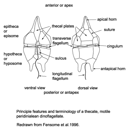
The patterning that you see is what I think to be the thecal plates. These fluoresce blue if you stain the specimen with calcofluor and can be another indication of Phylum Dinoflagellata

-
actinophrys

- Posts: 194
- Joined: Tue Oct 21, 2014 6:45 am
- Contact:
Re: Cylindrical Diatom
Spherical diatoms definitely do exist. Most form chains, as for instance in freshwater Melosira, but it looks like some also occur as single cells, like Podosira from seaweeds. Apparently auxospores can also be rounded, but I do not know much about how they look. In any case, though, I think the lovely detail in this photo – the look of the chloroplasts, hexagonally arranged punctae, and apparent flatness of the equatorial band – support it as some such diatom.
-
gconcepcion
- Posts: 14
- Joined: Mon Jan 07, 2019 11:39 pm
Re: Cylindrical Diatom
I don't necessarily disagree, what I was originally were thinking as theca are certainly the plastids (brushes off some of the rust...)... but it could still be a gymnodinoid (naked/athecate) dinoflagellete or another family. The punctae patterning also makes me think diatom, however dino's also show patterning at high magnifications: Dinoflagellate high resolutionactinophrys wrote:Spherical diatoms definitely do exist. Most form chains, as for instance in freshwater Melosira, but it looks like some also occur as single cells, like Podosira from seaweeds. Apparently auxospores can also be rounded, but I do not know much about how they look. In any case, though, I think the lovely detail in this photo – the look of the chloroplasts, hexagonally arranged punctae, and apparent flatness of the equatorial band – support it as some such diatom.