Inside Centropyxis
Posted: Tue Jun 23, 2020 5:42 pm
Hello Forum,
usually you observe disc shaped testate amoebae like Arcella or Centropyxis sitting flat on your glass slide, like this Centropyxis discoides:
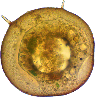
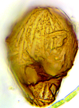
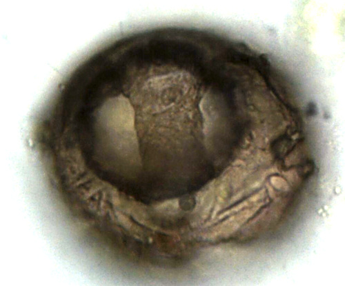
With some luck and glycerol I have been able to view the following two specimen from lateral. The next photos show some quite complex structural elements connecting dorsal and ventral shell, quite complex for a single-celler:
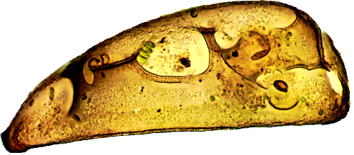
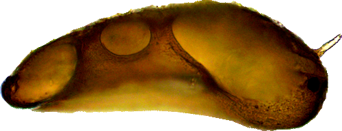
Diameters (from top) 240, 237 and 218 µm, Neofluar 40x, stacks of abt. 20 focal planes.
Cheers,
Hans
usually you observe disc shaped testate amoebae like Arcella or Centropyxis sitting flat on your glass slide, like this Centropyxis discoides:
With some luck and glycerol I have been able to view the following two specimen from lateral. The next photos show some quite complex structural elements connecting dorsal and ventral shell, quite complex for a single-celler:
Diameters (from top) 240, 237 and 218 µm, Neofluar 40x, stacks of abt. 20 focal planes.
Cheers,
Hans
 .
.