] So what does the long cylinder that sits above the field lens do? That confused me.[/quote]
I am not 100% sure Gekko but:
the glass below it , doesn't appear to be a lens, just a dust guard. That upper lens is centerable and appears to be focusable too, so it would seem to be where they have put the field lens. It has a filter tray on top too. Removing that would affect the settings and performance of the illumination system dramatically. Looking under the instrument , I can't see any way the collector lens is focusable.
Image quality of AmScope microscope via the trinocular tube
-
apochronaut
- Posts: 6400
- Joined: Fri May 15, 2015 12:15 am
Re: Image quality of AmScope microscope via the trinocular tube
I cannot disagree with apochronaut on the historical use of the field aperture to achieve even illumination.
The field aperture, if present, plays a large role in contrast and resolution with low power objectives. The proper use of the field aperture with low power obj. is to stop down the condenser aperture to the point of visibility in the edges of the field of view, then pull back a bit. Then stop down the field aperture to achieve the best contrast and resolution.
The above only applies to low power obj. and a microscope with a field aperture.
With high power obj. on modern microscopes with LED illumination or a halogen light source with a diffuser in the light path, the field aperture is used to control the shape of the illuminating cone.
At least that is the way it works on my Labomed LB-592.

Re: Image quality of AmScope microscope via the trinocular tube
Many thanks, apochronaut. That is very interesting. I take it you have one like Astyanax's? If so, do you think that the field iris assembly is "user replaceable" (I am thinking if Astyanax gets a "not working" S KE with a good iris)?apochronaut wrote:I am not 100% sure Gekko but:gekko wrote: So what does the long cylinder that sits above the field lens do? That confused me.
the glass below it , doesn't appear to be a lens, just a dust guard. That upper lens is centerable and appears to be focusable too, so it would seem to be where they have put the field lens. It has a filter tray on top too. Removing that would affect the settings and performance of the illumination system dramatically. Looking under the instrument , I can't see any way the collector lens is focusable.
I found an S-KT on ebay (looks rather similar, but I'm not sure if it uses the same field iris): http://www.ebay.com/itm/NIKON-BINOCULAR ... 257f4383fb (it appears to have the right eyepieces but wrong objectives).
I just looked at the Nikon site and found a picture of what they dsignate is S-Ke: http://www.microscopyu.com/museum/modelske.html It looks the same as Astyanax's SK E, but it doesn't have that cylindrical part, which suggests that in that version the field lens is at the level of the field iris control ring on the base. Puzzling. Well, I just read the description which confirms what you said, that the cylinder contains the field lens-- I guess the picture shows it without the field lens assembly: "The S-Ke was the first Nikon microscope equipped with a Köhler illuminator built into the microscope base, enabling convenient and perfect photomicrography when a camera was mounted and used with one of Nikon's Microflex adapters. A reflecting mirror was provided as an accessory and could replace the illumination field lens to provide light from a source outside the microscope.'
Focusable collector lens: In my Nikon Optiphot, the collector lens is also not focusable: the whole lamp housing (bulb and collector lens) is moved as a unit to focus the filament. My rather vague memory of my work microscope (Zeiss IM) is that the collector was focusable.

Re: Image quality of AmScope microscope via the trinocular tube
Thanks apochronaut and gekko. Apochronaut's last comment is spot on about the washer. it sits about a mm higher than the original diaphragm and yes backing off the condenser is practical and does not make much difference.
I will take on board your suggestions to stick with the Nikon. There is a body of one from eBay for ~$US200 delivered. But who knows what condition the thing is. I'll try to contact the seller and find out. Certainly having a working iris is useful though.
gekko thanks for confirming my thinking about the reduction lens. It's not a big expense may be worth getting one to try out.
Any comments from other colleagues who may have used a 0.4x or a 0.3x reduction lens would be welcome.

Re: Image quality of AmScope microscope via the trinocular tube
I think that gekko's suggestion of an external illuminator is a practical and effective solution, if perhaps a little more unwieldy than the present arrangement.
As is also the case in the external illuminator from Carl Zeiss Jena shown.
"The S-Ke was the first Nikon microscope equipped with a Köhler illuminator built into the microscope base, enabling convenient and perfect photomicrography when a camera was mounted and used with one of Nikon's Microflex adapters. A reflecting mirror was provided as an accessory and could replace the illumination field lens to provide light from a source outside the microscope.'
In my case, the Lamp House (Zeiss 60w illuminator) and the collector lens are fixed - the bulb is moved to focus the filament.Focusable collector lens: In my Nikon Optiphot, the collector lens is also not focusable: the whole lamp housing (bulb and collector lens) is moved as a unit to focus the filament. My rather vague memory of my work microscope (Zeiss IM) is that the collector was focusable.
As is also the case in the external illuminator from Carl Zeiss Jena shown.
Last edited by 75RR on Mon Jun 15, 2015 8:59 pm, edited 1 time in total.
Zeiss Standard WL (somewhat fashion challenged) & Wild M8
Olympus E-P2 (Micro Four Thirds Camera)
Olympus E-P2 (Micro Four Thirds Camera)
Re: Image quality of AmScope microscope via the trinocular tube
I posted the pictures below. I also give below my first visual impressions looking through the microscope and at the camera's LCD, but you can now evaluate the pictures.
I will try to write this tediously precisely so that it would be difficult to misunderstand what I did.
1. All tests were done with the 4x objective and the diffusing filter, located in front of the condensing lens at the rear of the microscope in place. The images are numbered to correspond to the numbers given to the descriptions below. The image resolution was reduced from a width of 4032 pixels to 1024 pixels (the maximum size allowable for uploading to Photobucket, and for displaying on this forum).
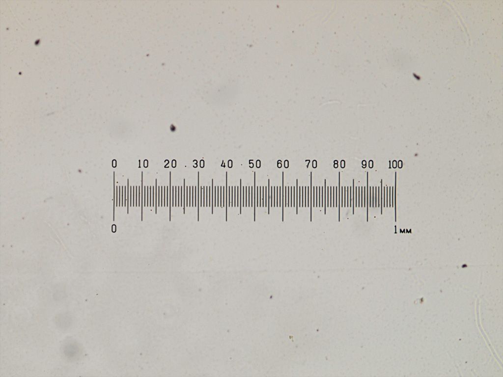
2. As I mentioned earlier, and as apochronaut indicated, and as The QCC I think said above, my Achromatic condenser with the flip-down top lens does not allow Koehler illumination, but my understanding is that it is similar to your condenser with the removable top lens. I set up the microscope as recommended by Nikon, namely, set up Koehler using the 10x objective and the top condenser lens in. Then, with the 4x in place, I flipped down the top lens without altering the condenser height. With the top lens flipped down, the condenser iris is left wide open, and the field iris acts as an aperture. I used it in this configuration. I adjusted the field iris to get about 85% of the objective aperture filled with light. My impression is that the uniformity of illumination was at least acceptable. The image is of my stage micrometer, which is at the same scale as the images of the slide below.
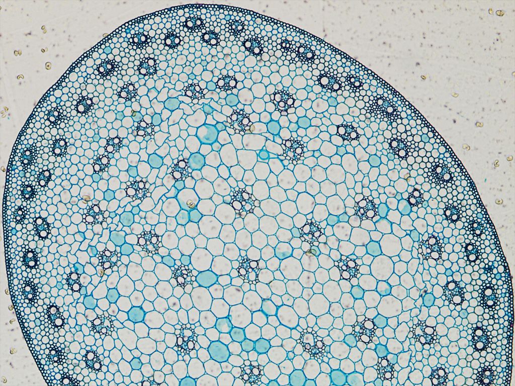
3. I used the Achromatic condenser with the top flipped down, and the microscope adjusted as in step 2 above and imaged a commercially prepared slide labeled "Stem of monocotyledon" (stained transverse section), with the condenser iris fully open and the field iris, which now acts as an aperture, illuminating about 85% of the 4x objective.
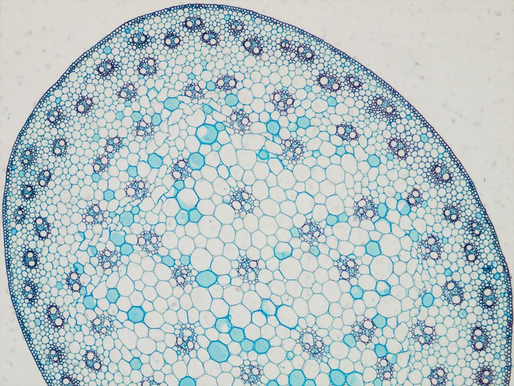
4. I repeated 3 above, but with the aperture (field iris) fully open to simulate your broken iris. My expectation is significant glare and loss of contrast.
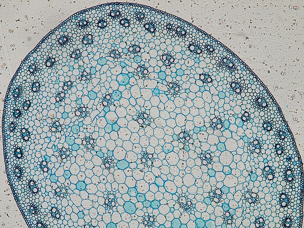
5. I repeated 4 above, but added on top of the field lens, an iris removed from an old condenser (I'll call that generic iris), adjusted to about 85% aperture. Since the field iris in the configuration using the flip-down condenser is essentially an aperture iris, I wanted to see if situating the generic iris on top of the field lens (with the microscope's field iris fully open) would give results similar to 3 above. The edge of the generic iris as viewed at the back focal plane of the objective appeared to my eye as sharp as the microscope's field iris even though it was much closer to the condenser and on the other side of the field lens. My visual impression of the image produced was very favorable and not visibly different from that obtained under 3 using the field iris as an aperture (i.e. normally adjusted microscope with a good field iris). This I think simulates your microscope with the condenser top lens removed, and with an iris obtained from a cheap old condenser placed above the field lens, using the 4x objective. If it truly does simulate that, then I think you are in business. However, your field lens is much closer to the bottom of the condenser than mine is, and I don't know what the effect of that would be. You can easily test that by removing the top condenser lens, and then placing something (paper clip, pencil, whatever) just above the field lens (so you don't scratch it) while observing the back focal plane of the objective to see if what you put above the field lens is in focus at the objective's back focal plane.

6. I repeated 5 above, but with the generic iris fully closed (to show that it does indeed act pretty much like an aperture diaphragm).
I will try to write this tediously precisely so that it would be difficult to misunderstand what I did.
1. All tests were done with the 4x objective and the diffusing filter, located in front of the condensing lens at the rear of the microscope in place. The images are numbered to correspond to the numbers given to the descriptions below. The image resolution was reduced from a width of 4032 pixels to 1024 pixels (the maximum size allowable for uploading to Photobucket, and for displaying on this forum).
2. As I mentioned earlier, and as apochronaut indicated, and as The QCC I think said above, my Achromatic condenser with the flip-down top lens does not allow Koehler illumination, but my understanding is that it is similar to your condenser with the removable top lens. I set up the microscope as recommended by Nikon, namely, set up Koehler using the 10x objective and the top condenser lens in. Then, with the 4x in place, I flipped down the top lens without altering the condenser height. With the top lens flipped down, the condenser iris is left wide open, and the field iris acts as an aperture. I used it in this configuration. I adjusted the field iris to get about 85% of the objective aperture filled with light. My impression is that the uniformity of illumination was at least acceptable. The image is of my stage micrometer, which is at the same scale as the images of the slide below.
3. I used the Achromatic condenser with the top flipped down, and the microscope adjusted as in step 2 above and imaged a commercially prepared slide labeled "Stem of monocotyledon" (stained transverse section), with the condenser iris fully open and the field iris, which now acts as an aperture, illuminating about 85% of the 4x objective.
4. I repeated 3 above, but with the aperture (field iris) fully open to simulate your broken iris. My expectation is significant glare and loss of contrast.
5. I repeated 4 above, but added on top of the field lens, an iris removed from an old condenser (I'll call that generic iris), adjusted to about 85% aperture. Since the field iris in the configuration using the flip-down condenser is essentially an aperture iris, I wanted to see if situating the generic iris on top of the field lens (with the microscope's field iris fully open) would give results similar to 3 above. The edge of the generic iris as viewed at the back focal plane of the objective appeared to my eye as sharp as the microscope's field iris even though it was much closer to the condenser and on the other side of the field lens. My visual impression of the image produced was very favorable and not visibly different from that obtained under 3 using the field iris as an aperture (i.e. normally adjusted microscope with a good field iris). This I think simulates your microscope with the condenser top lens removed, and with an iris obtained from a cheap old condenser placed above the field lens, using the 4x objective. If it truly does simulate that, then I think you are in business. However, your field lens is much closer to the bottom of the condenser than mine is, and I don't know what the effect of that would be. You can easily test that by removing the top condenser lens, and then placing something (paper clip, pencil, whatever) just above the field lens (so you don't scratch it) while observing the back focal plane of the objective to see if what you put above the field lens is in focus at the objective's back focal plane.
6. I repeated 5 above, but with the generic iris fully closed (to show that it does indeed act pretty much like an aperture diaphragm).
Last edited by gekko on Tue Jun 09, 2015 1:34 am, edited 2 times in total.
Re: Image quality of AmScope microscope via the trinocular tube
Thanks, 75RR. I would interpret that to be the same as a focusable collector lens, in the sense that the distance between the filament and the collector lens is adjustable. In the case of my Nikon Optiphot, that distance is fixed. Very nice lamp, by the way.75RR wrote:In my case, the Lamp House (Zeiss 60w illuminator) and the collector lens are fixed - the bulb is moved to focus the filament.
Re: Image quality of AmScope microscope via the trinocular tube
For $200 delivered, especially if it is the same model, it looks like a very good deal, even to use for parts. But if it is in good working conditions, I would jump at it.Astyanax wrote:Thanks apochronaut and gekko. Apochronaut's last comment is spot on about the washer. it sits about a mm higher than the original diaphragm and yes backing off the condenser is practical and does not make much difference.
I will take on board your suggestions to stick with the Nikon. There is a body of one from eBay for ~$US200 delivered. But who knows what condition the thing is. I'll try to contact the seller and find out. Certainly having a working iris is useful though.
gekko thanks for confirming my thinking about the reduction lens. It's not a big expense may be worth getting one to try out.
Any comments from other colleagues who may have used a 0.4x or a 0.3x reduction lens would be welcome.
Just to reiterate about the reduction lenses, as apochronaut and I mentioned above, they do not provide the requisite optical corrections for your Nikon objectives, so they will not provide the best images, especially for 10x and higher objectives (the larger the NA the more obvious the effect). However, I think that with the 4x, they will be fine.
Re: Image quality of AmScope microscope via the trinocular tube
If this is the Microsope you are talking about at $US200 delivered - I would go for it if I were you.
http://www.ebay.com/itm/Complete-Nikon- ... 33a2b1f14e
http://www.ebay.com/itm/Complete-Nikon- ... 33a2b1f14e
Zeiss Standard WL (somewhat fashion challenged) & Wild M8
Olympus E-P2 (Micro Four Thirds Camera)
Olympus E-P2 (Micro Four Thirds Camera)
Re: Image quality of AmScope microscope via the trinocular tube
I agree with 75RR if this is what you were looking at. If not, I would get this one. Shipping to Australia is $58. It comes with a full complement of objectives, which alone would be worth the cost. I would get it before it goes. Seller has 100% feedback.
Re: Image quality of AmScope microscope via the trinocular tube
Thanks 75RR Unfortunately that was not the one I looked at that one is an S. The one I saw was a base only of precisely my model S-KE but it is no longer listed. Looks like its gone 
gekko - wow! you have done a lot of work I can hardly wait to see your pics. The field lens (tube on the scope foot) comes out and at the base there is a glass base/filter of some kind which can be unscrewed. As it happens it sits ~ a mm away from the original broken diaphragm. So a thin rubber washer works well. Of course it is a nuisance having to change it for different objectives. As you said having it fully open there is too much glare etc. I'll try your idea on top of the field lens if I can find a derelict iris. In theory it should work since I can see the surface scratches of the filed lens in focus when the system is set (washer edge in focus).
By the way you may be interested to have a look at the link I found. The SKe is described in pretty nice detail along with the base condenser's settings functions. (http://www.microscopy-uk.org.uk/mag/ind ... nikon.html).
I'm going to take a few pics myself shortly. (Just waiting for the temp to go up a bit. The scope is in the garage and while Melbourne winter is not as severe as yours its pretty nippy!)
Re: Image quality of AmScope microscope via the trinocular tube
It was fun and I learned something. I posted the images "in-line" in my previous post.Astyanax wrote:gekko - wow! you have done a lot of work I can hardly wait to see your pics.
Re: Image quality of AmScope microscope via the trinocular tube
The images are GREAT - Actually I prefer the one simulating the fully open situation.
You have done a fantastic job with the different settings.
I tried a few simulations with the washer but I think as you correctly pointed out my field lens is much closer to the condenser and the washer is not in focus when on top of the field lens. But the bottom position seems best. However, viewing looks much better than photos - old story!
The results are disappointing - terrible in fact.
Even with the 10x set with top lens in (focus on washer below the field lens) there is unevenness second pic. To add insult to injury my power supply spat the dummy and I was only able to run at the the lowest light intensity level. Sorry my book keeping is not as meticulous as yours but you will get the idea. It was not worth posting the rest of it.
First pic is with a 10x obj, second 4x obj - scale and the 4x slide (both with the top condenser lens off)[filament centred and focused]. The same unevenness is evident at normal light level settings before the p/s malfunction. The unevenness can be seen clearly even slight out of focus. Just cannot even it out. The low light level tends to accentuate it but it was evident before the p/s spat the dummy. (The phantom parallel lines are a function of the low light level camera interaction. The camera was not running at base I had the gain turned up to about 50%, where normally I run the camera at minimal gain (least noise) and adjust the light level.
That is starting to look like a new microscope is needed!
- Attachments
-
- IS1a.jpg (119.14 KiB) Viewed 12061 times
-
- IS8.jpg (106.1 KiB) Viewed 12065 times
-
- IS10aa.jpg (68.61 KiB) Viewed 12065 times
Re: Image quality of AmScope microscope via the trinocular tube
In fact it shouldn't be visible at all. With top lens of the condenser removed, the washer is acting as an aperture, so it should not be visible in the eypeices or camera, but should be focused at the back focal plane of the objective, so you should be able to see it if you remove an eyepiece and look down at the back of the objective.Astyanax wrote:the washer is not in focus when on top of the field lens.
I think you should separate unevenness of illumination from a bad image (low resolution), and should separate the quality of the image through the eyepieces from that of the camera. For example if the image looks good in eyepiece but bad in the camera, it is not the microscope's problem.
Unevenness of illumination: is it tht the center is brighter than the edge? Or is it that one side is brighter than the opposite side? The images of the wood section suggest to me that the left side is darker than the right. The image of the stage micrometer, however, suggests, if anything, that the edges are slightly darkened but looks to me fairly evenly illuminated At least that is what I see. The unevenness from one side indicates to me that there is something obstructing the light from one side, but that whatever is obstructing the light is not there when you use your stage micrometer. Or am I missing something?
Unevenness aside, are you happy with the resolution you obtained looking down the microscope? Same question looking at the camera photos?
Thank you! So, if you prefer the one with fully open field iris, simulating your broken field iris, then you can use your microscope as is [without a field iris] (that is, assuming my simulation actually does indeed simulate as intended).Astyanax wrote:The images are GREAT - Actually I prefer the one simulating the fully open situation.
Re: Image quality of AmScope microscope via the trinocular tube
This issue of there appearing to be something in the way has always been the problem. It was a while since I last took lots of images with the scope and I am starting to forget about old problems. Resolution is not an issue but uniformity and edge falloff both of illumination and focus is. Its as if there is a problem with the alignment of the phototube even though all the connections/links are secure. When the filament is perfectly centered and focused on the bottom of the condenser iris its good for viewing through the eyepieces but it appears to deviate through the phototube (the same through an eyepiece instead of the camera, so its unlikely to be a function of the camera) I have even attempted to recentre the filament but there is just no adjustment that works. Sticking the camera into one of the viewing tubes instead overcomes the problem albeit still with a small focal reduction at the edges. I'll try to redo some more pics through the viewing tube once I get the p/s working.
Re: Image quality of AmScope microscope via the trinocular tube
I don't see a lever to switch that directs all the light to the eyepieces or to the phototube. Is the light permanently directed partly to the eyepieces and partly to the phototube? If there is a lever to switch the light path, then it may be that it is not going all the way, so the phototube light path is partially obstructed? If there is no mechanical switch/lever, then the phototube may not be properly centered, or there is some obstruction in the head below the phototube? The question remains as to why the image of the stage micrometer does not show the unevenness (at least to my eyes).
Re: Image quality of AmScope microscope via the trinocular tube
Thanks gekko, there is a slider at the back. I considered that also tried switching all the light or partial and positioning to no avail.
-
apochronaut
- Posts: 6400
- Joined: Fri May 15, 2015 12:15 am
Re: Image quality of AmScope microscope via the trinocular tube
..it would be good to be getting a proper iris diaphragm in that thing. cobbled together microscopes are inevitably annoying. i was beginning to wonder when the duct tape was going to be unveiled.
look straight down the photo tube on low illumination( apparently that's all you have right now?) and move that slider back and forth. the light pathways on many older microscopes that were not designed for wider fields(20+), are quite restrictive and any little misalignment could cause enough blockage to give a shadowing effect.....and it not necessarily will affect the visual field. as well, look down the tube with a bright flashlight and the illuminator off. i have had instances where a very slight misalignment caused a vague shadow along part of the field and another where an old spider web was in there, making all kinds of trouble. Nikon , unusually did not have a direct straight through path from the objective to the photo tube. It is diverted, so there could be something in the head causing the problem or simply murk on the components.
also, low illumination shows up every imperfection and dust speck as blocked light, so the evenness of the field looks worse than it really is. if you can get your trafo. up to speed, you may see a dramatic improvement but I suspect your optical path to the photo tube needs attention.
are you sure your microscope is an SK-E? there was an SKT and an LK-E. One of the differences is in the arrangement of the optics in the base, with the SKT, having the nose extension above the base but normally in base electronics, with a switch and rheostat on the left side. The SK-E , I have not seen with the type of illuminating system you have. It was simpler, with the field lens back towards the bulb. They built these , along with the LK-E during a period when the electronics were being moved into the base, so you see two similar but different field lens extensions on them and some with a switch on the side. Your microscope looks to be an LK-E, virtually identical to the SKT but with a remote trafo.
Re: Image quality of AmScope microscope via the trinocular tube
Found a catalogue that may clarify model type. Modular microscopes can be a challenge.are you sure your microscope is an SK-E? there was an SKT and an LK-E.
http://micromundos.com.br/manuals/Nikon ... 201976.pdf
Zeiss Standard WL (somewhat fashion challenged) & Wild M8
Olympus E-P2 (Micro Four Thirds Camera)
Olympus E-P2 (Micro Four Thirds Camera)
-
apochronaut
- Posts: 6400
- Joined: Fri May 15, 2015 12:15 am
Re: Image quality of AmScope microscope via the trinocular tube
There you go . Great find!
Looks like an SK-E. I guess the differences are that the LK-E has 70mm of focus travel and a 5 hole nosepiece. It does show the light path for the photo tube. Seems the light has to pass through a beamsplitter, for a 30/70 light division and also can be routed through a prism for 0/100. This moving prism could be murking up the photo image. The other trinocular is an either or prism but it doesn't look like the one on this scope.
Looks like an SK-E. I guess the differences are that the LK-E has 70mm of focus travel and a 5 hole nosepiece. It does show the light path for the photo tube. Seems the light has to pass through a beamsplitter, for a 30/70 light division and also can be routed through a prism for 0/100. This moving prism could be murking up the photo image. The other trinocular is an either or prism but it doesn't look like the one on this scope.
Re: Image quality of AmScope microscope via the trinocular tube
Thanks 75RR. Good to have the catalogue.
apochronaut:
I think you have hit the nail on the head! Using a variable dc power supply I was able to drive the filament lamp and looked at the result. There is still the same shadowing through the photo tube, not that evident viewing by eye but they still shows up in the pics. Using slides with good contrast (strong stain) it becomes less of an issue but its still not right. However, with low contrast it is easily visible.
I suspect your first point may right that there may be a design restriction. Alternatively, but less likely, that there is crud on the prism. If so I can't see it - there is no difference after cleaning. There is also a round filter sitting in a small well which can be seen when the head is removed. This is an analyser as for a Polarized work and it has always been there. taking it out makes no difference. I normally leave it in there. It's occasionally useful to stick a polariser in the bottom path for spotting crystals within cells or for determining specific orientations of cell types.
I will try to take a few more pics through one of the observation tubes. Initially that seemed provide a better result.
-
apochronaut
- Posts: 6400
- Joined: Fri May 15, 2015 12:15 am
Re: Image quality of AmScope microscope via the trinocular tube
One thing I have seen more than I would like to have in old mics., are small areas of cement separation in cemented prisms or beam splitters. Often you cannot see it unless the you hold a light just so. That could be causing the small area of unevenness, even though the prisms look clean. Hopefully not, because the only solutions are radical; decementing and recementing the prism or replacement from a junker( but not too much of a junker) OR if the prism is toast anyway, a cheap and sometimes good fix is to immerse it in 100% ethyl alcohol. If the cement is Canada Balsam, this most likely will work. You have to time it right and drain the alcohol just as the cement slightly dissolves and reforms. The trick is to put the prism into a container with a lid, just big enough to hold it. Cover with alcohol, then lid. When pickled enough, gently take off the cap, hold the prism down with your index finger and pour the alcohol off. leave alone for 24 hours. If it is synthetic cement, which I suspect it is, the same could be done by knowing the cement type and the solvent. Immersing in ethyl alcohol won't do anything, so you can always try , if you don't know the type of cement. I've done it a couple of times with yes and no success. Probably not best to try it though, unless you have decided 100% that it needs to be replaced anyway.
Re: Image quality of AmScope microscope via the trinocular tube
Thanks apochronaut for the method but every time I touch stuff it gets buggered up. I wouldn't dare disassemble the head.
As a final trial, I stuck the camera down the observation tube instead of the photo tube. Result is a tad better, but the condenser setting as per conventional K. setting (without the field iris) with the condenser top lens on, in this case, seems to be the best. See the pics. There is a small central bright spot, did not see it before taking the pics but it's there, no big deal, its barely visible under normal observation.
First image 10x, rest 4x, and scale with a blue filter. Have to get a power supply.
I think its time I gave up and look to get a newy for decent micrography.
- Attachments
-
- IS4.jpg (97.58 KiB) Viewed 11975 times
-
- IS.jpg (134.16 KiB) Viewed 11975 times
-
- IS8.jpg (24.51 KiB) Viewed 11975 times
Re: Image quality of AmScope microscope via the trinocular tube
I can see the attraction of a quick and in all probability a perfectly adequate solution.I think its time I gave up and look to get a newy for decent micrography.
Perhaps it is time to find out what "with a 1yr warranty" means.To cut a long story short. TechTrader is offering the XY-Series scope with alternate plan optics: 4, 10, and 40x less an oil immersion 100x for $Au1000.00 delivered to Melbourne. The scope is their shop demo unit but they are still offering it with a 1yr warranty (whatever that means).
At that price you might want to try to get them to throw in a 20x or 25x
Zeiss Standard WL (somewhat fashion challenged) & Wild M8
Olympus E-P2 (Micro Four Thirds Camera)
Olympus E-P2 (Micro Four Thirds Camera)
Re: Image quality of AmScope microscope via the trinocular tube
I empathize with your frustration. I think you've tried your best to save it. What I am not sure is what an xy-series is (who makes it)? How does it compare, say, with microscopes offered by microscope.net or microscopeworld in terms of qulaity, price, warranty, etc.
http://www.microscopeworld.com/
http://www.microscopenet.com/?gclid=CIe ... gQodkEwA8Q
I must add that I've never dealt with either company nor do I know anything about them, but they seem to be microscope 'speacialists' in the sense of that is what they sell, but again, I don't know who TechTraders are. Keep us updated with what you decide to do and best of luck!
Re: Image quality of AmScope microscope via the trinocular tube
It would appear that the XY Series is made by Sunny Optical Technology, a relatively new company (2004).
Its main business seems to be making lenses for mobile phone cameras.
http://www.sunnyoptical.com/en/009005002/p389.html
Its main business seems to be making lenses for mobile phone cameras.
http://www.sunnyoptical.com/en/009005002/p389.html
Zeiss Standard WL (somewhat fashion challenged) & Wild M8
Olympus E-P2 (Micro Four Thirds Camera)
Olympus E-P2 (Micro Four Thirds Camera)
Re: Image quality of AmScope microscope via the trinocular tube
Thanks 75RR and gekko
After several calls and exchange of emails I got a couple more images from the scope from TechTrader.
The scope they were offering is a shop demo model they were not able to move and originally outfitted for phase (not plan) they were going to replace the objectives with inf. plans not sure what kind. But I was not happy with the images and their price crept up too. An additional problem was the fastness of the plastic components, they were becoming sticky to the touch this had to be removed which made the scope look tatty - anything but new. I was not impressed with the dirty lenses either. In the end I think they were getting sick of the questions and and put me on the spot to buy the scope. I declined.
So I am back to square 1.
I cheked out the OMAX's from your links gekko. They look ok, but interestingly they don't seem to have units with both Koehler illum. and plan optics; its one or the other. The Koehler's don't look elaborate, all I can see is that they just seem have a field iris. However, to get a Koehler unit I have to buy another set of plan objectives and throw away the achros and most Koehler models have small stages....
Thus far I am still edging towards the T720/Amscope except it comes with infinity objectives which is of limited flexibility, i.e., I can't use my good Nikon plans in case the the supplied objectives are "ordinary"
By the way I cleaned the SKe as best as I could and it is great for observation now.
As a final point I wonder if it is worth getting one of the 0.3x reduction lenses to try with my system (~$Au65 not a great cost). If that should give me a larger field of view maybe I can crop off the edges and still get a reasonable image at low mag?
In any case I will keep you posted.
Re: Image quality of AmScope microscope via the trinocular tube
Might well be for the best. A company that sells second hand microscopes at such outrageous prices (AU$ 4000+) is unlikely to be an honest broker.So I am back to square 1.
I was thinking about your comments on the Infinity Amscope or indeed any Amscope (they are not all infinity), as in what do you get that you do not already have (if you get that stand on ebay) and the answer is just the Trinocular and if you get yours working properly again not even that!
Last edited by 75RR on Fri Jun 12, 2015 6:29 pm, edited 1 time in total.
Zeiss Standard WL (somewhat fashion challenged) & Wild M8
Olympus E-P2 (Micro Four Thirds Camera)
Olympus E-P2 (Micro Four Thirds Camera)
Re: Image quality of AmScope microscope via the trinocular tube
Double post. Sorry
Zeiss Standard WL (somewhat fashion challenged) & Wild M8
Olympus E-P2 (Micro Four Thirds Camera)
Olympus E-P2 (Micro Four Thirds Camera)