#10
Post
by charlie g » Tue Nov 07, 2017 3:27 am
Again, thanks for the specifics on your new SONY with electronic shutter, and high ISO performances,hkv (thanks, Jim T for asking about this).
I suggested 1.5% methylcellulose viscosity tool as..you the microscopist do not function at: ISO of 1/2000 th's. You scan an elongated droplet of your sample on a slide with no applied cover slip...this easy with 4X, 10X, even 20X objectives. When you determine you have target highly mobile organisms to observe...you plop drops of methylcellulose to the wetmount...and then place the rectangular coverslip on this prep. You only feed waters to such a wetmount over the time you then use the slide...not more methycellulose.
You can not track high speed organisms for use of 1/2000 image captures ( unless there is a microscopy Aegis missle system tracking coupled to the mechanical stage, I sense.)...I really appreciate your beautiful and crisp microscopy shared experiences, hkv, I would never suggest awkward placement of methylcellulose to the edge of a coverslip-wetmount prep.
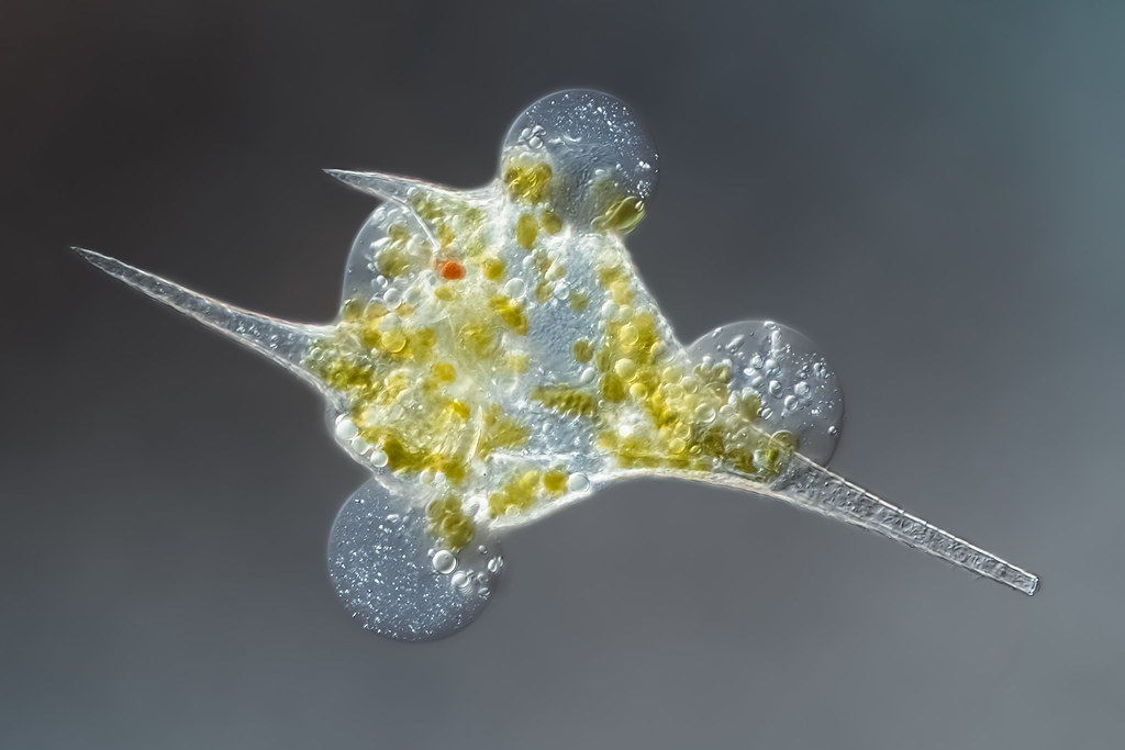
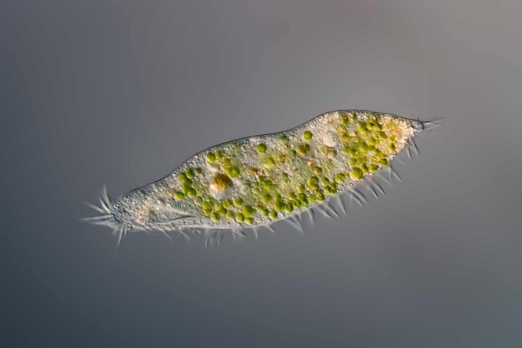
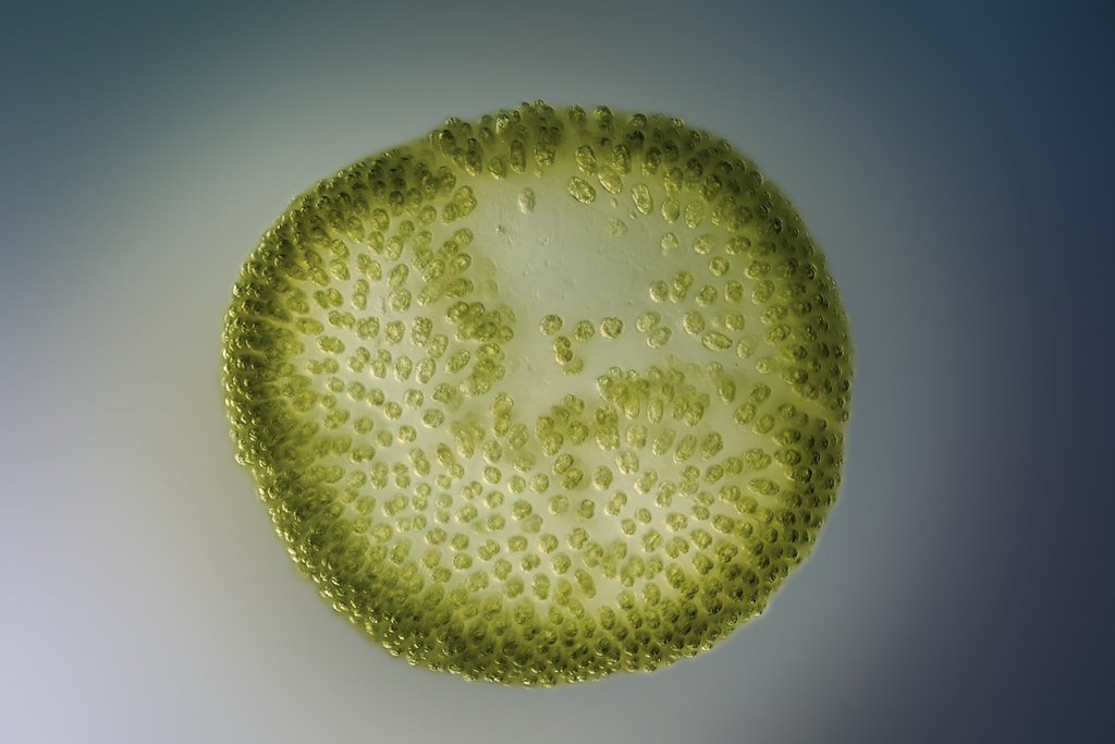
Thank you,hkv for these beautiful protozoan images. Charlie guevara