Have problems identifying an organism? Ask for help here.
-
Wes
- Posts: 1027
- Joined: Sat Mar 09, 2019 12:58 pm
#1
Post
by Wes » Sun Aug 04, 2019 8:46 am
Last edited by
Wes on Sun Aug 04, 2019 11:26 am, edited 3 times in total.
Zeiss Photomicroscope III BF/DF/Pol/Ph/DIC/FL/Jamin-Lebedeff
Youtube channel
-
tgss
- Posts: 223
- Joined: Wed Jan 25, 2017 3:48 am
- Location: Ontario, Canada
#2
Post
by tgss » Sun Aug 04, 2019 11:17 am
I'm afraid I could only see one of the four images Wes. (The fourth)
Tom
-
Wes
- Posts: 1027
- Joined: Sat Mar 09, 2019 12:58 pm
#3
Post
by Wes » Sun Aug 04, 2019 11:27 am
Thanks for letting me know, Tom. Does it work now?
Zeiss Photomicroscope III BF/DF/Pol/Ph/DIC/FL/Jamin-Lebedeff
Youtube channel
-
Crater Eddie
- Posts: 1858
- Joined: Wed Nov 12, 2014 4:39 pm
- Location: Illinois USA
#4
Post
by Crater Eddie » Sun Aug 04, 2019 12:12 pm
I see all four. No idea what it is though.
CE
Olympus BH-2 / BHTU
LOMO BIOLAM L-2-2
LOMO POLAM L-213 / BIOLAM L-211 hybrid
LOMO Multiscope (Biolam)
Cameras: Canon T3i, Olympus E-P1 MFT, Amscope 3mp USB
-
Wes
- Posts: 1027
- Joined: Sat Mar 09, 2019 12:58 pm
#6
Post
by Wes » Sun Aug 04, 2019 12:49 pm
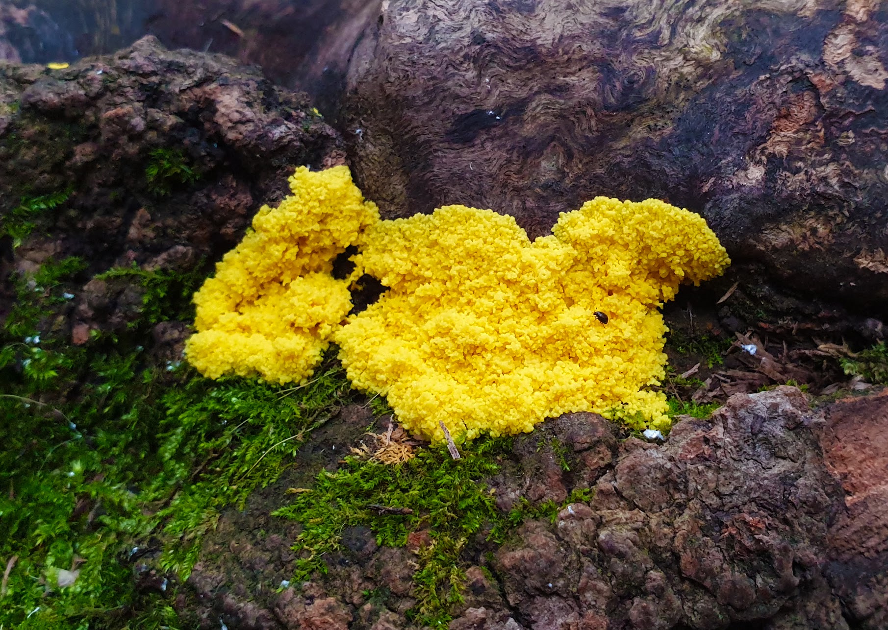
After some browsing it looks very similar to Fuligo septica also known as
dog vomit slime mold 
I have it in a petri dish now, on a piece of wood surrounded by a wet paper towel and another piece on agar with bacteria. Will keep an eye on it for a few days, maybe something interesting like sporulation happens.
Zeiss Photomicroscope III BF/DF/Pol/Ph/DIC/FL/Jamin-Lebedeff
Youtube channel
-
tgss
- Posts: 223
- Joined: Wed Jan 25, 2017 3:48 am
- Location: Ontario, Canada
#7
Post
by tgss » Sun Aug 04, 2019 1:42 pm
Well, as the previous posts have made clear, it works fine now, for me as for everyone else

No idea what it is, though it's certainly slime mouldish in appearance. The first picture showing the macroscopic view is quite spectacular though.
Tom
-
MLTechie
- Posts: 2
- Joined: Sun Nov 03, 2019 12:36 pm
#8
Post
by MLTechie » Sun Nov 03, 2019 12:54 pm
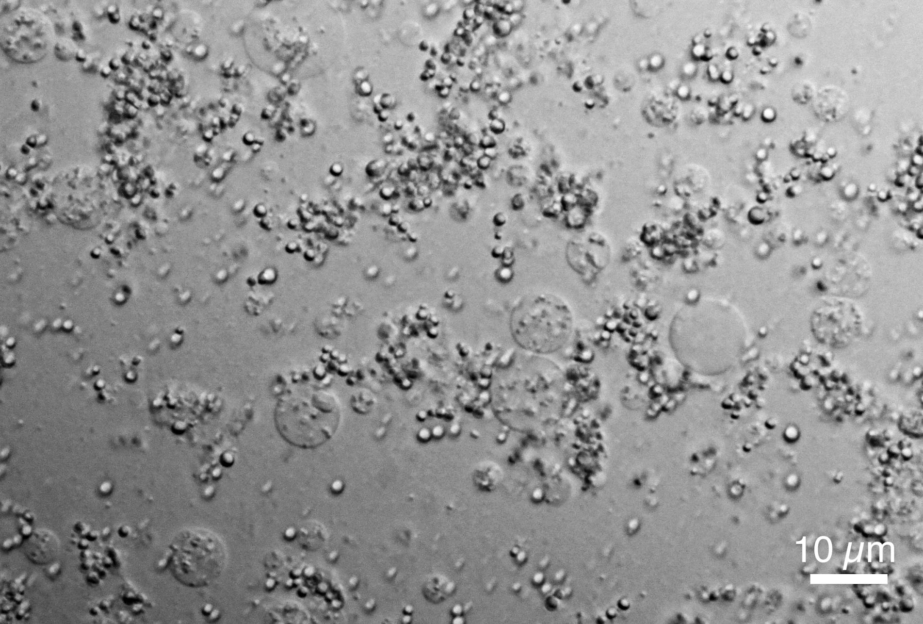
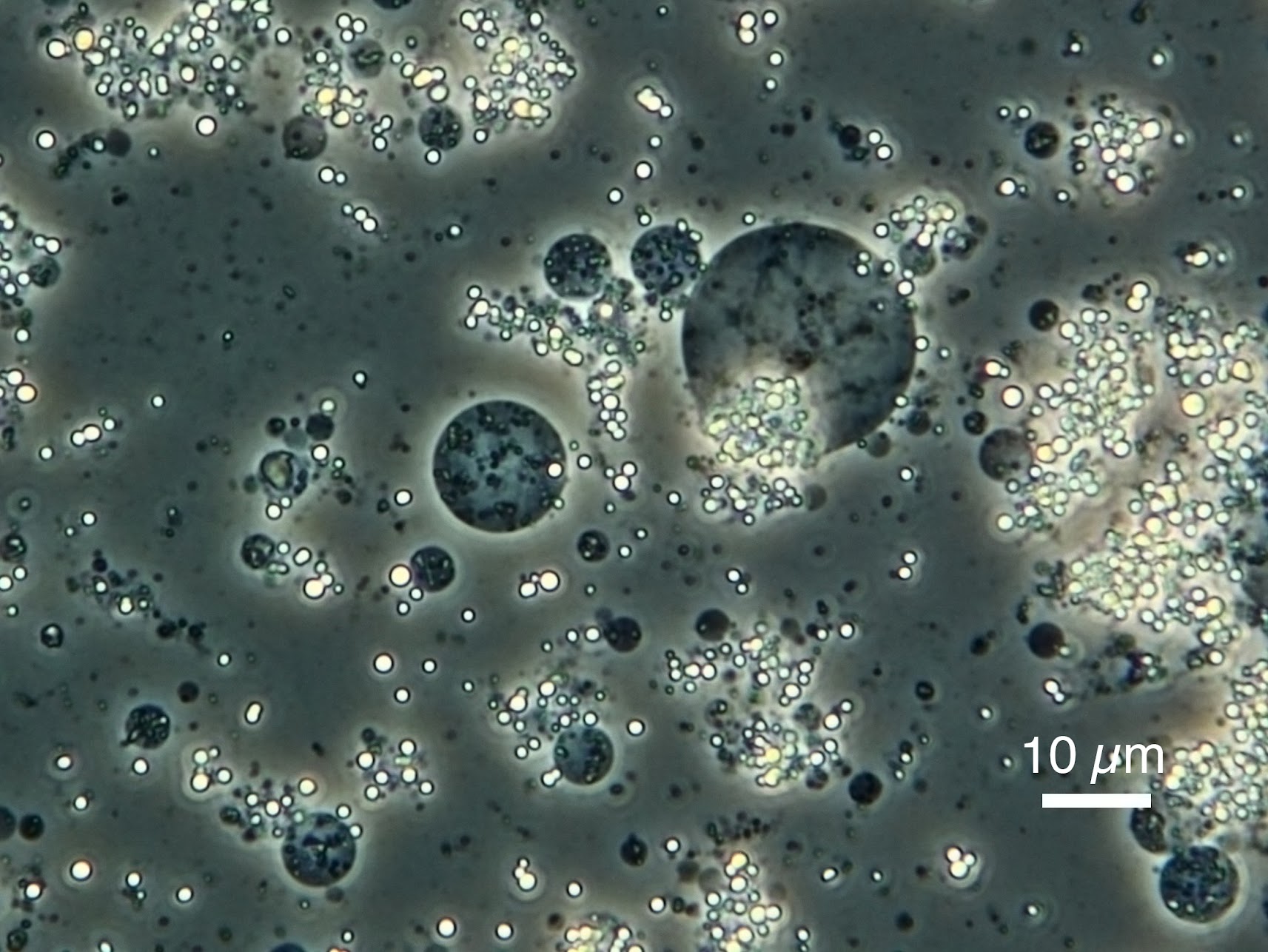
I concur! It appears to be the spore phase of a slime mold, as you mentioned it is some fuligo spp..
Check this link from research gate, it shows the life cycle.
https://www.researchgate.net/figure/Lif ... _242179415
The thing is, it is difficult to collect anything but the spores unless you grow it in a Petri dish. We normally do a tease mount and LPCB stain for fungi in my class. If you could find the swarm cells then it would be even more specific (if you want to really Narrow it down more to be a myxomycete).
Thanks for sharing,
MLTechie
-
mnmyco
- Posts: 144
- Joined: Tue Aug 28, 2018 11:03 pm
#9
Post
by mnmyco » Sun Nov 03, 2019 10:48 pm
It is definitely a slime mold and not a Tremella species or any other fungus.