Page 1 of 1
making longitudinal sections
Posted: Tue May 25, 2021 10:44 am
by iconoclastica
I have been making transverse sections through the stalks of leaflets of ferns (below). Now I would like to demonstrate that the reddish brown cells are fibers really. For this I need to make sections perpendicular to the one below, i.e. following the length of the stalk. My present microtome doesn't seem cut out for this purpose. Are there any suggestions how to make longitudinal sections?

- D. x complexa 10x methylene blue
- D complexa 10x-1.jpg (63.05 KiB) Viewed 6518 times
Re: making longitudinal sections
Posted: Tue May 25, 2021 6:40 pm
by mrsonchus
Hi, I had a quick look at these cells this afternoon in some fern in my garden. I took some very basic longitudinal sections (hand) and had a look over them....
They do surround the vascular-strand of the fern stem/leaf/leflet but I'm not sure they are actually 'fibers' as defined for flowering plants - in fact I haven't had time yet to look further into it.
Here are a few very basic water-mounted hand-section images in LS but they don't really add much info I'm afraid, perhaps the simple-pitting of the cell-walls is helpful?


The fern-leaf tissue may be an interesting subject to make into proper sections.....
Re: making longitudinal sections
Posted: Tue May 25, 2021 6:42 pm
by BramHuntingNematodes
Embedding may be called for.
Re: making longitudinal sections
Posted: Tue May 25, 2021 6:57 pm
by DonSchaeffer
It may seem primative, but I make logitudinal sections by essentially scraping layers from the plant with a rzor-knife. I have been doing it under the stereo microscope. It usually leaves a sample 2 or 3 cell layers thick but you can focus through the transparent cells.
Re: making longitudinal sections
Posted: Tue May 25, 2021 7:16 pm
by MicroBob
Hi,
there is a very simple method:
Fresh plant material contains water, which has the effect to harden cyanacrylate glue. Just prepare a block of wood and glue the fresh material onto it. When sectioning stay away from the glue with the knife as it would dull the knife quickly. Do the fixation after sectioning.
Bob
Re: making longitudinal sections
Posted: Tue May 25, 2021 8:19 pm
by iconoclastica
mrsonchus wrote: ↑Tue May 25, 2021 6:40 pm
Hi, I had a quick look at these cells this afternoon in some fern in my garden. I took some very basic longitudinal sections (hand) and had a look over them....
You seem to have taken a sample from
Dryopteris filix-mas that indeed does not have the additional sclereids. What your section shows is one of the two round "eyes" in my section. It's a vascular-strand with a sheath of sclrerified cells around it. Where they branch off, the spiral-ringed vessels can be beautifuly seen.
Your remark on the pitting of the cell walls intrigues me. I have been observing and photographing them for the past hours, but I didn't know how to interprete them further than 'probably pits'. Is simple pitting a special type of it? My botany labs have been too long ago...
DonSchaeffer wrote: ↑Tue May 25, 2021 6:57 pm
It may seem primative, but I make logitudinal sections by essentially scraping layers from the plant with a rzor-knife.
Don, would that be real scraping (with the knife perpendicular to the surface) of cutting with the knife in an angle?
MicroBob wrote: ↑Tue May 25, 2021 7:16 pm
there is a very simple method:
Fresh plant material contains water, which has the effect to harden cyanacrylate glue. Just prepare a block of wood and glue the fresh material onto it.
Thanks Bob, that certainly is one to try. I was going to make a variant of my microtome this week, with a slit rather than a hole, and I imagine that could work very well.
Re: making longitudinal sections
Posted: Tue May 25, 2021 9:55 pm
by DonSchaeffer
Roughly perpendicular.
Re: making longitudinal sections
Posted: Wed May 26, 2021 12:24 am
by mrsonchus
Hmm, looking at your fine image closer does suggest an endodermis, as the thickened walls look to have the typical pattern of an endodermis as in the roots of angiosperms....
In fact a quick search seems to confirm that the strands each have their own endodermis - the thickenings of the inner-tangential and vertical anticlinal (radial may be a better term?) cell-walls is just about visible in your TS image. What view in particular are you looking for?
Simple pits are just straight channels through (secondary) cell-walls whereas other forms such as 'bordered' pits have different structures such as a chamber before the innermost primary cell-wall is reached. In answer to your question yes simple pits are one a several types of pit found in plant cell-walls, the presence of which allows communication/transport through thickened secondary cell-walls and of course the primary cell-walls either side of the middle lamella of two adjacent cells.
Re: making longitudinal sections
Posted: Wed May 26, 2021 9:19 am
by iconoclastica
I may not have been clear enough about what I am looking at. In the image below I have highlighted the sclereids of interest:

- D complexa 10x-1.jpg (55.25 KiB) Viewed 6432 times
Detail of centre with vascular bundle and a few sclereids. Some of the latter shows a transverse cell wall with what I interpreted as pits:

- xcomp vasculis 40x.jpg (210.35 KiB) Viewed 6432 times
The most detailed I can get it:
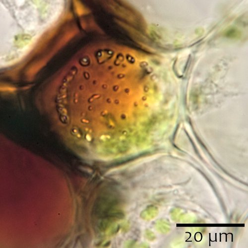
- sectio costae 100x_0100.jpg (47.64 KiB) Viewed 6432 times
Re: making longitudinal sections
Posted: Fri May 28, 2021 7:33 pm
by iconoclastica
MicroBob wrote: ↑Tue May 25, 2021 7:16 pm
there is a very simple method:
Fresh plant material contains water, which has the effect to harden cyanacrylate glue. Just prepare a block of wood and glue the fresh material onto it.
This indeed works excellently. I made a new version of my microtome, one that allows for flat objects to be cut too. I glued the stalk to a strip of cardboard and got results on the first try:
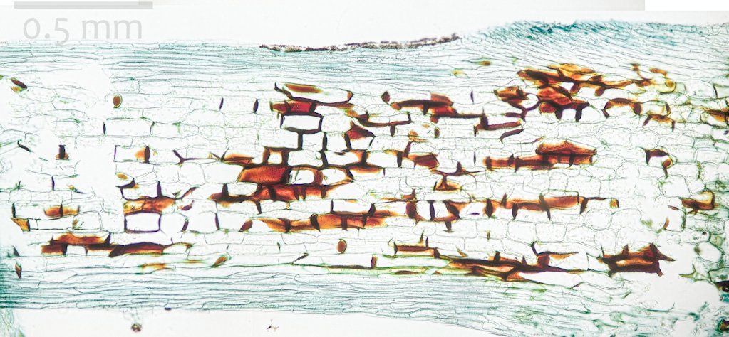
- longitudinal section of the pinnae stalk and costa (10x)
- affinis sectio longitudinalis 10x.jpg (148.34 KiB) Viewed 6370 times
It's immediately clear that the red-brown cells are not typical fibers: not spindel shaped and not very long. I tend to call it 'collenchymatous', but I hope someone here has better knowledge of plant anatomy than I have...
Re: making longitudinal sections
Posted: Fri May 28, 2021 7:42 pm
by iconoclastica
Within the series of longitudinal sections, the veins got amost tangentially sectioned, allowing great views on the transporting tissue:

- vein with endodermis (brown) and tracheids (20x)
- affinis tracheids 20x.jpg (54.25 KiB) Viewed 6370 times
The tracheids come in various sizes:

- tracheids, some with spiral thickenings (40x)
- affinis tracheids 40x 2.jpg (69.5 KiB) Viewed 6370 times
and their typical endings can be seen, although with my .90 condensor I couldn't well see the end plates:

- tracheids continuing into the next ones (100x)
- affinis tracheids 100x 3.jpg (70.62 KiB) Viewed 6370 times
Finally, pits in the endodermis cell walls:

- endodermis with pits (40x)
- affinis pits 40x.jpg (42.09 KiB) Viewed 6370 times
Re: making longitudinal sections
Posted: Fri May 28, 2021 11:03 pm
by tgss
Very good results. Well done, and thanks for sharing.
Tom W.
Re: making longitudinal sections
Posted: Sat May 29, 2021 11:41 am
by perrywespa
Great images. Being fairly new to sectioning, I'd like to see a picture of the specimen glued to the cardboard, if possible. Thanks!
Re: making longitudinal sections
Posted: Sat May 29, 2021 7:40 pm
by iconoclastica
perrywespa wrote: ↑Sat May 29, 2021 11:41 am
Great images. Being fairly new to sectioning, I'd like to see a picture of the specimen glued to the cardboard, if possible. Thanks!
I don't have a picture of that, but here's the reconstruction:
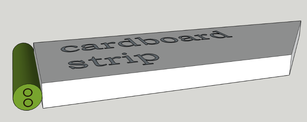
- Untitled.png (61.49 KiB) Viewed 6090 times
I dipped the short end into a blob of cyano-acrylate glue and pressed it onto the stem fragment.
Re: making longitudinal sections
Posted: Tue Jun 08, 2021 10:02 pm
by perrywespa
Thanks, that's kind of what I thought. I may try that this weekend. Sorry for the long response time.