
1. Oblique illumination:
2. DIC:
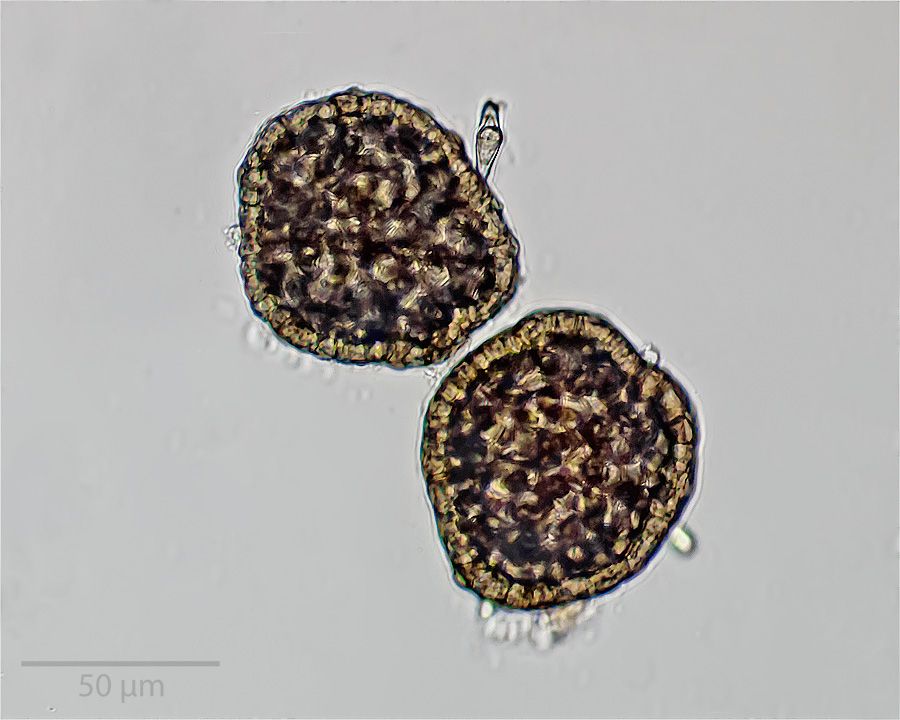
Thank you, seb, for your suggestion. I don't think it is amoeba for the reasons I mentioned above in my reply to 75RR (what look like "pseudopods" did not move, it was flat with thickness about 10 µm, and focusing through it did not show any structure inside other than what we see in the images).vasselle wrote:Bonjour Gekko
ça ressemble fort un amibe.
Cordialement seb
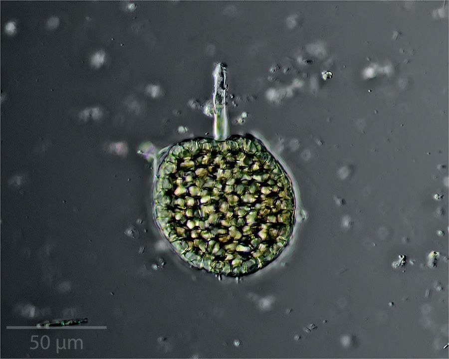
I think Peter is right and we are looking at a beautiful micrograph of a germinating pollen grain.gekko wrote:Peter, thank you for your suggestion. I guess they could be pollen, but does the fact that they are flat (please see my answer to 75RR above) or that they have "stems" (most of the ones that I saw in the field of view appeared to have stem-like projections) argue against them being pollen? I don't know enough to say one way or the other.
Had a thought, whereas pollen is, as far as I know always, prompted into 'doing something' by the addition of water - 'plumping-up' and often growing a pollen-tube (this can happen very quickly and may be 'watched live'), spores tend to 'do something' when they are dried/dry - that is their capsule (that which we may be seeing in your pictures) will not only split open from a special area (stomium) of thinner cells, but peel-back as a result of a specialized structure (annulus) that often looks a lot like a 'spine' contracting then 'pinging' the contents of the capsule (the spores themselves) outwards. Try letting a droplet of liquid (water?) containing these objects simply desiccate upon a slide then see if you 'get a reaction'.... Might be very interesting..gekko wrote:Many thanks, John, for your input. I tend to agree with you in that all the pollen that I've seen is spherical or nearly so, and those are quite flat and, therefore, what you say makes sense to me, despite my ignorance. Perhaps they are some kind of spore.
Yes, I would say it resembles my picture perhaps more than a little. That, to my eyes (bad as they are) is the closest thing I've seen. Regarding your suggestion to desiccate them, well I've seen those things only that one time, but if I ever see them again, that would be what I would do and see what happens. Very useful new (to me), interesting, and useful information in your reply. Thank you for taking so much trouble and also sharing so much expertise and information!mrsonchus wrote:Had a thought, whereas pollen is, as far as I know always, prompted into 'doing something' by the addition of water - 'plumping-up' and often growing a pollen-tube (this can happen very quickly and may be 'watched live'), spores tend to 'do something' when they are dried/dry - that is their capsule (that which we may be seeing in your pictures) will not only split open from a special area (stomium) of thinner cells, but peel-back as a result of a specialized structure (annulus) that often looks a lot like a 'spine' contracting then 'pinging' the contents of the capsule (the spores themselves) outwards. Try letting a droplet of liquid (water?) containing these objects simply desiccate upon a slide then see if you 'get a reaction'.... Might be very interesting..gekko wrote:Many thanks, John, for your input. I tend to agree with you in that all the pollen that I've seen is spherical or nearly so, and those are quite flat and, therefore, what you say makes sense to me, despite my ignorance. Perhaps they are some kind of spore.
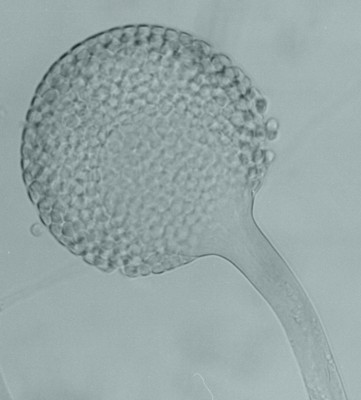
Here's a picture - not all annuli are as obvious as this one - some are zonal areas rather than the classic 'backbone' type that is very obvious in this picture..
https://classconnection.s3.amazonaws.co ... 808532.jpg
The center chamber (containing the spores) resembles your picture a little.


