Vaginicola

Here you can post pictures and videos to show others.
Post Reply
Message
Author
molehill
Posts: 30
Joined: Wed Jun 24, 2015 10:24 am

Vaginicola

#1 Post by molehill » Mon Jun 20, 2016 10:31 am

It is now mid winter in Auckland, New Zealand. Even though the weather has been very mild I was a bit disappointed
with the lack of activity in the water sample I collected from a stream which runs through the park behind our house.
I found Stentor in this stream earlier on in the year. However it wasn't completely lifeless. .
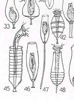
I looked through a old book to give me a clue. A google search turned up a meagre but useful result.

Vaginacola ........ I love the old print

Also notice the banding in the phase image.

The images where made with a Meiji Techno MX5000 and a Canon EOS 40D


Image

Image
Meiji S Apo 20x 0.65 400ASA 1/250sec

Image
Meiji U Plan PH 20x 0.40 400ASA 1/20sec

User avatar
rnabholz
Posts: 3086
Joined: Thu Jan 08, 2015 10:11 pm
Location: Iowa USA
Contact:

Re: Vaginicola

#2 Post by rnabholz » Mon Jun 20, 2016 11:29 am

Sorry your sample wasn't more active, but what you did get is really interesting!

I had never seen this before, and you present two great shots of it, and that super vintage illustration. Thanks for sharing and the introduction.

User avatar
75RR
Posts: 8207
Joined: Sun Oct 12, 2014 2:34 am
Location: Estepona, Spain

Re: Vaginicola

#3 Post by 75RR » Mon Jun 20, 2016 11:32 am

However it wasn't completely lifeless. .
How true ... Excellent catch!
Zeiss Standard WL (somewhat fashion challenged) & Wild M8
Olympus E-P2 (Micro Four Thirds Camera)

billporter1456
Posts: 492
Joined: Thu Oct 16, 2014 12:05 am

Re: Vaginicola

#4 Post by billporter1456 » Mon Jun 20, 2016 12:35 pm

Nice images and I loved the old illustration you provided. Vaginicola is one of my favorite organisms to photograph. Thanks for sharing!

User avatar
zzffnn
Posts: 3206
Joined: Sat Jun 20, 2015 3:57 am
Location: Houston, Texas, USA
Contact:

Re: Vaginicola

#5 Post by zzffnn » Mon Jun 20, 2016 1:21 pm

Very beautiful photos, thank you!

User avatar
exmarine
Posts: 247
Joined: Mon May 09, 2016 1:16 am
Location: Cambridgeshire UK

Re: Vaginicola

#6 Post by exmarine » Mon Jun 20, 2016 2:15 pm

Great photo's and now a little bit about this Protozoa. This is a loricated peritrich ciliate. The cytoplasm of this species is green owing to the symbiotic green algae. The ciliates project from the lorica when feeding but can contract into it for protection when stimulated e.g. by vibration.
It is not uncommon for two cells to inhabit the same lorica.

Many larger species like Vaginicola have the ability to contract , a device which protects them from turbulence in the surrounding water, or from larger predators, (flatworms and snails) which may glide over the submerged material.

I find its always good to know something about these protozoa.
Thank you :shock:
Best regards
exmarine :x

uses Watson 'Service' 1950 compound.
uses Watson Stereo 1960 ish.

JimT
Posts: 3247
Joined: Fri Oct 24, 2014 1:57 pm

Re: Vaginicola

#7 Post by JimT » Mon Jun 20, 2016 2:20 pm

It is now mid winter in Auckland
Stay warm :)

Very nice images and the accompanying illustration and thank you Exmarine for the additional info.

User avatar
gekko
Posts: 4701
Joined: Sun Oct 12, 2014 7:38 am
Location: Durham, NC, USA.

Re: Vaginicola

#8 Post by gekko » Mon Jun 20, 2016 2:37 pm

Great to see your beautiful images again, molehill. It is nice that you show the peritrich extended as well as contracted. Excellent photos. The brightfield captures delicate details with great clarity. The phase contrast image has its own beauty, and nicely reveals the bands in the lorica that remain invisible in brightfield (I've never seen those before). Do you, by any chance, have a phase contrast image with the Vaginicola extended?

billporter1456
Posts: 492
Joined: Thu Oct 16, 2014 12:05 am

Re: Vaginicola

#9 Post by billporter1456 » Mon Jun 20, 2016 4:17 pm

gekko wrote:The phase contrast image has its own beauty, and nicely reveals the bands in the lorica that remain invisible in brightfield (I've never seen those before).
I am wondering if the "bands" might be an artifact. I done lots of video of Vaginicola with phase contrast and never seen those bands. These two videos are examples: [/youtube]

Maybe some interaction between the illumination (LED?) and the camera? I certainly don't know for sure, just wondering.

Bruce Taylor
Posts: 1023
Joined: Fri Sep 04, 2015 11:34 am

Re: Vaginicola

#10 Post by Bruce Taylor » Mon Jun 20, 2016 7:39 pm

molehill wrote: I love the old print
I recognize it. :) A few years ago, I cropped out and enhanced the ciliate images from C. G. Ehrenberg's Die Infusionsthierchen als vollkommene Organismen and posted them in a couple of places on the net. That pic of Vaginicola tinctus was one of them.

Lovely pics of a sweet organism...thanks for showing them here! I especially like the phase contrast image, which shows the faint annuli of the lorica, may be left over from the construction process. Donald Ott, an algologist at U. of Akron, has made a rather wonderful time-lapse video which shows how Cothurnia (a closely related ciliate) builds its little bottle-home:

https://www.youtube.com/watch?v=6J_L0ntoDrY

billbillt
Posts: 2895
Joined: Mon Nov 10, 2014 10:01 pm

Re: Vaginicola

#11 Post by billbillt » Mon Jun 20, 2016 8:05 pm

I took the liberty of downloading and enlarging an area of these "bands"... It appears they are individual rings as Bruce Taylor points out.. They must be the segments that make up the lorica..
Attachments
bands.jpg
bands.jpg (78.44 KiB) Viewed 6490 times

billporter1456
Posts: 492
Joined: Thu Oct 16, 2014 12:05 am

Re: Vaginicola

#12 Post by billporter1456 » Mon Jun 20, 2016 9:26 pm

Bruce Taylor wrote: I especially like the phase contrast image, which shows the faint annuli of the lorica, may be left over from the construction process.
Hi Bruce, thanks for chiming in on the "bands." Strange that I have never seen them despite doing several phase contrast videos of Vaginicola and Cothurnia. But if they were an illumination artifact, I guess they would have extended across the field of view.

Bruce Taylor
Posts: 1023
Joined: Fri Sep 04, 2015 11:34 am

Re: Vaginicola

#13 Post by Bruce Taylor » Mon Jun 20, 2016 11:53 pm

Bill, in the phase contrast view on your first video -- around 0:57 - 1:00 or so -- I imagine I can see very faint annuli on the right side of the lorica...a series of faint horizontal shadows...

Or, maybe I'm nuts. :D

In any case, there is a fair bit of variation among (and within!) species. Distinct annular corrugations are fairly common on vaginicolid loricae (and in many other ciliates, such as Metacystis or any number of tintinnids), so perhaps it isn't so strange if subtle "bands" are visible in some "hyaline" loricae but not others. Some Cothurnia from Kahl:

Image

billporter1456
Posts: 492
Joined: Thu Oct 16, 2014 12:05 am

Re: Vaginicola

#14 Post by billporter1456 » Tue Jun 21, 2016 12:20 am

Bruce Taylor wrote:Bill, in the phase contrast view on your first video -- around 0:57 - 1:00 or so -- I imagine I can see very faint annuli on the right side of the lorica...a series of faint horizontal shadows...

Or, maybe I'm nuts. :D

In any case, there is a fair bit of variation among (and even within!) species. Distinct annular corrugations are fairly common on vaginicolid loricae (and in many other ciliates, such as Metacystis or any number of tintinnids), so perhaps it isn't so strange if subtle "bands" are visible in some "hyaline" loricae, but not others. Some Cothurnia from Kahl:

Image
Bruce, thanks for that great information! It's really nice to have someone as knowledgeable as you on the forum to help out. And while you may be "nuts," you are correct about the faint shadows in my YouTube video. I just went back to the original video file and looked at it closely. The horizontal shadows (6 or 8 of them) are definitely there on the right side of the lorica.

User avatar
zzffnn
Posts: 3206
Joined: Sat Jun 20, 2015 3:57 am
Location: Houston, Texas, USA
Contact:

Re: Vaginicola

#15 Post by zzffnn » Tue Jun 21, 2016 12:24 am

Bruce Taylor wrote:Donald Ott, an algologist at U. of Akron, has made a rather wonderful time-lapse video which shows how Cothurnia (a closely related ciliate) builds its little bottle-home:

https://www.youtube.com/watch?v=6J_L0ntoDrY
Thank you, Bruce, for your comments.

It is great to see a pro's work. Dr. Ott has lots of high quality and interesting YouTube videos, in addition to many research publications.

User avatar
gekko
Posts: 4701
Joined: Sun Oct 12, 2014 7:38 am
Location: Durham, NC, USA.

Re: Vaginicola

#16 Post by gekko » Tue Jun 21, 2016 1:11 am

I'd like to also add my thanks to Bruce. As I think I said before, every time Bruce (or for that matter "actinophrys") writes something, we learn something.

Edit: corrected typo.
Last edited by gekko on Tue Jun 21, 2016 12:58 pm, edited 1 time in total.

molehill
Posts: 30
Joined: Wed Jun 24, 2015 10:24 am

Re: Vaginicola

#17 Post by molehill » Tue Jun 21, 2016 11:04 am

Thank you all for the lively discussion, links, videos and analysis of my post, very much appreciated.
It is a great way to learn.
Unfortunately I didn't get a phase image of the Vaginicola fully extended, I was concentrating on the banding
So I hope to find some more specimens in the water sample I have.
I noticed the lorica in billporters video is almost transparent whereas in my image it is optically a lot denser then
the surrounding medium. Could this account for the banding showing up more prominent in my phase image?

Molehill

Bruce Taylor
Posts: 1023
Joined: Fri Sep 04, 2015 11:34 am

Re: Vaginicola

#18 Post by Bruce Taylor » Tue Jun 21, 2016 11:17 am

zzffnn wrote:.It is great to see a pro's work. Dr. Ott has lots of high quality and interesting YouTube videos, in addition to many research publications.
If you need to know everything about Vaucheria, he's your man. :D A really nice guy, too...which seems to be a common trait among microscope enthusiasts, professional and amateur alike.

Post Reply