apochronaut wrote: ↑Mon Jun 01, 2020 12:02 pm
great images. When highly polished images show up on this forum, it is always a question of how much the final presented image relates to the viewed image , at the point the image was captured. Most microscopists spend 99.9% of their microscope time viewing through the microscope and perhaps the rest capturing and or processing images, if that. A lot of questions on this forum and I also see on the more dedicated photo...forum are about how to present better photos, the answer to at least some of which is in the post processing. I know even my simple pictures can be improved a lot with some fiddling. Cleaning the sensor once in a while for instance. Would there be any chance of showing us one or a few of the original image captures of these, so we can get an idea of how much the p.p. has affected the final outcome?
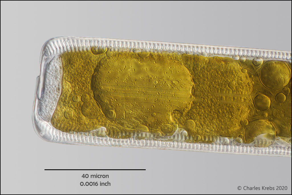
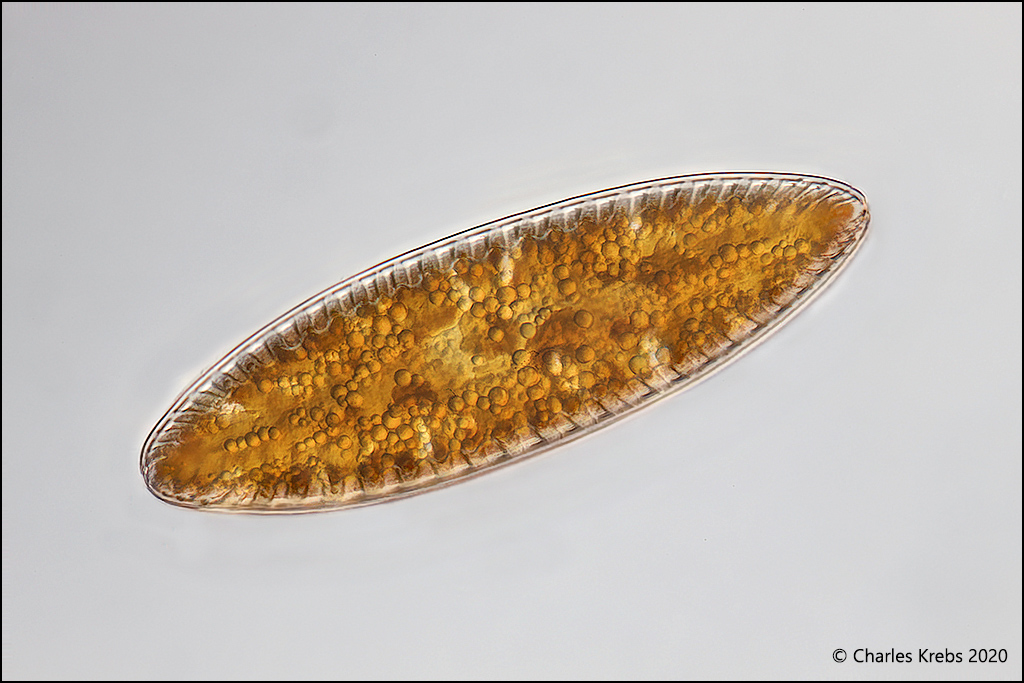
Good questions. As a kid, my first microscope "project" was making a collection of pollen slides which I photographed through the eyepiece with a clunky Polaroid B+W camera. (I hate to think about it, but that was about 60 years ago!). Put the microscope away after a few years, but stuck with photography. I got "hooked" again when digital photography became viable... in large part because of the ability to tackle the nemesis of crazy shallow DOF by using image stacking. The images here, and frankly the vast majority of images I post are focus stacked. While I am very much aware of the pitfalls of stacking, I feel that the final images (can) present a far more satisfying view of the subject than a single frame that has perhaps 1/100th the DOF. (And if there is any question about detail representation I still retain all the "raw" camera files that can be scrutinized if need be). When you examine a subject visually under the microscope, you are usually constantly tweaking the fine focus to put the section you are looking at in best focus. So there is much less of a sense inadequate DOF as when you look at a single image still photograph. Looking at a high magnification photograph (single frame, not stacked), especially one made with a high NA objective can be frustrating for a viewer since they don't have the ability to turn the fine focus and see detail in sections where they would like to.
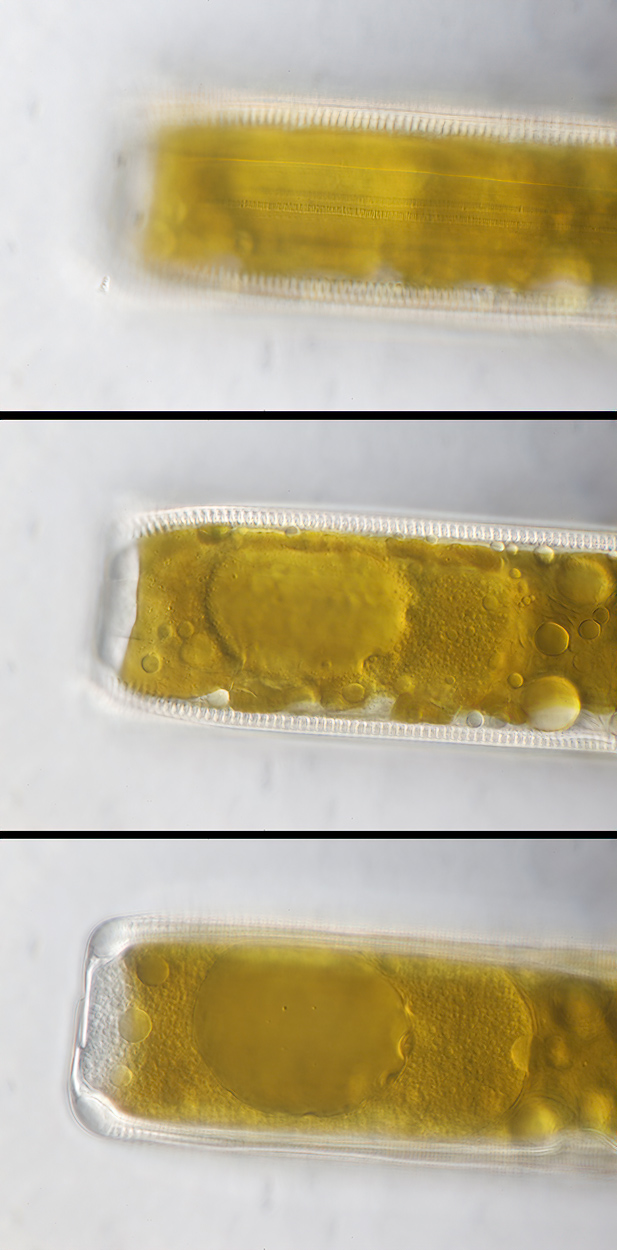
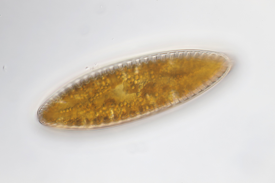
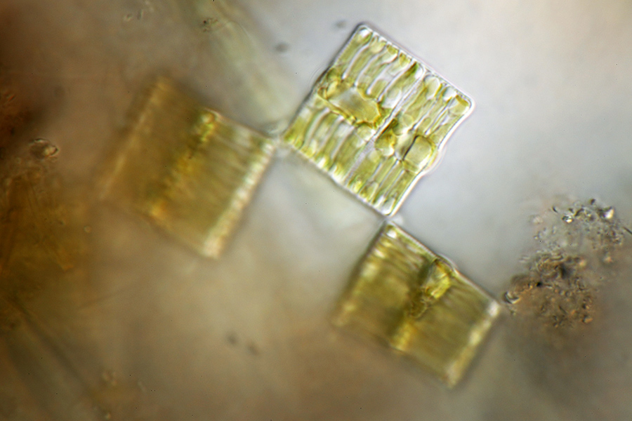
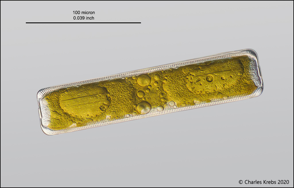
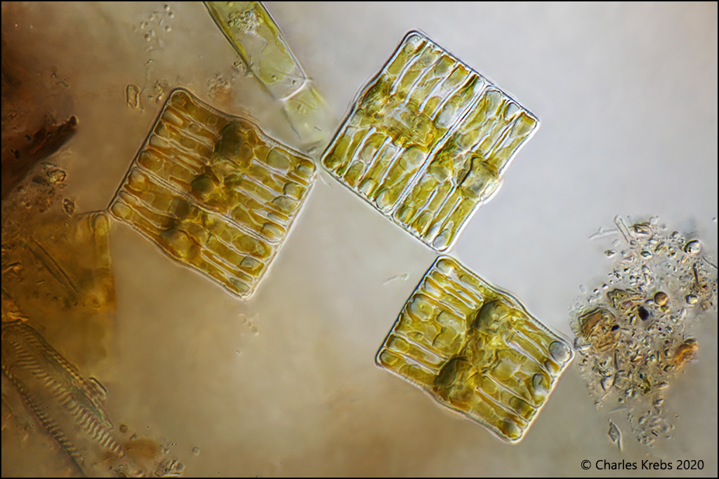
Focus stacking is major "post processing". The resulting image will usually require some digital cleanup. Sometimes a great deal of it. For example, in the third image above the diatom was moving (rotationally and laterally). The stacking software (Zerene in this case) did an admirable job of aligning the layers. But that means that every speck of dust on the sensor, every little bit of debris on the slide get repeated in the background in a "track" mimicking the diatoms motion. (For the first three images above, I was very meticulous in selecting the individual diatoms and placing them in a very clean wet mount with distilled water, so I avoided the normal "debris". They are seldom this clean). Below I have put some of the original "source" files. In the first triptych you can see one from the beginning of the stack, one from the middle and one from near the end. To me, none of these individual frames is completely satisfying if I want to see the entire subject photographed. The stack is (Image #2 above). I have also included single frames from the other images originally posted. Again (to me at least) not as interesting or effective as the stacks.
But this is not just an argument for stacking. With digital we have the ability to present images without the air bubbles, coverslip or slide defects, fibers and unrelated debris that made it into the mount. I guess a purist might say that's all part of the reality. But if it is foreign to the subject and adds no useful context (it rarely does), I don't like it

.
And there is overall correct color balance, proper tonality. (I find it fascinating to look through some of the old 'microscope photography" texts to see all the techniques film photographers needed to use to accomplish what can be done now with some basic post-processing. The "pre-processing" in those days was often quite daunting!) Slide preparation is rarely perfect. The images that any camera spits out (film or digital) are not perfect renditions of the scene before it. If you have an interest in presenting microscope images to people, you are, IMO, doing yourself and your viewers a disservice if you do not have at least some understanding and capability of basic post-processing.