P-protein strand from tobacco

Here you can post pictures and videos to show others.
Post Reply
Message
Author
CaterpillarGuy
Posts: 85
Joined: Thu Jul 09, 2015 3:08 am

P-protein strand from tobacco

#1 Post by CaterpillarGuy » Sun Jul 19, 2015 3:36 am

I thought you guys might get a kick out of seeing an occasional TEM shot from ~40 years ago. No one else in my life has ever been interested in seeing this. ;-) It's from my Master's project in bio at UC Santa Barbara.

Image

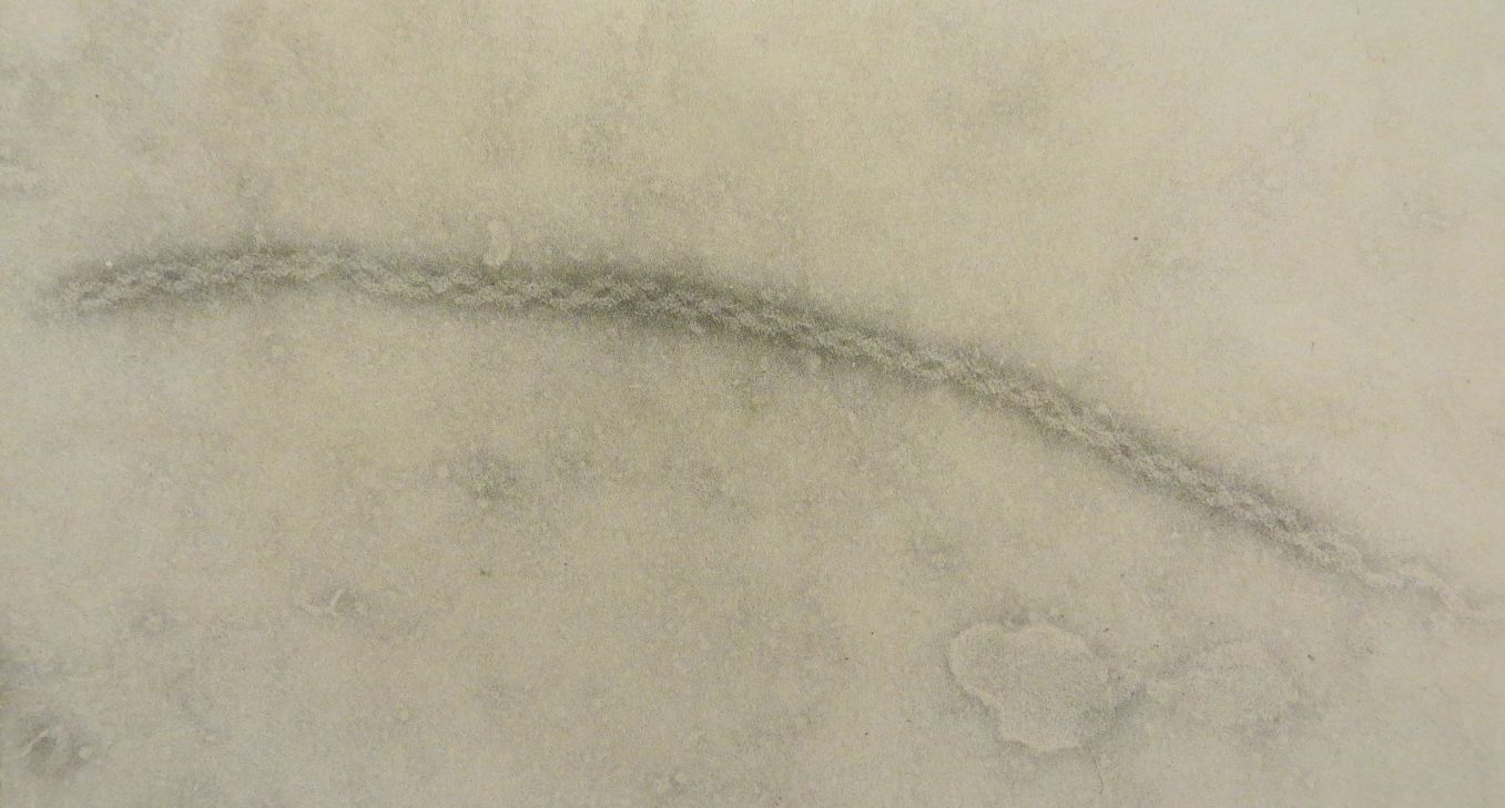
This is an image of a negatively-stained "P-protein" strand (P = phloem) from a tobacco (Nicotiana) plant. I have had this print sitting around since about 1973 because it was my favorite shot of these proteins. Really easy to shoot pics of these: Slice a young tobacco plant in mid stem, and over a minute or so a liquid exudes from the cut stem, much of it coming from the phloem. The exudate is collected in a pipette, diluted, and a tiny drop squirted onto a coated TEM grid (coated with a very thin film of sputtered carbon) and stained with a solution of electron-dense phosphotungstic acid. Remove excess stain via edge of Kimwipe, allow the grid to dry for a minute or two, stick it in the scope and spend half an hour looking around the grid at fairly high mag. Just a few minutes from intact plant to electron beam, which is VERY nice compared with the usual multi-day tedium of prepping TEM specimens for sectioning.

Pretty cool helix, no? The diameter (~20nm or ~200 angstroms) is similar to that of a microtubule. Magnification on my print was 135K and the diameter of the structure measured 5mm on the print, so you can use that to see what the mag is on your computer screen.

The scope was a brand-new one of these: http://www.science.oregonstate.edu/emfa ... y/EM/EM300 and is now probably considered worthy of Smithsonian display. ;-)

Edit: "negative" staining means that the stain (phosphotungstic acid) settles in around the biologic structure rather than staining the structure itself. All the dark areas are stain, and the light areas are relative absence of stain, and the pattern of stain reveals the structure of the protein. Biological tissues/structures aren't dense enough to deflect electrons and hence are transparent in a TEM; so in any TEM (transmission EM) image, anything dark in the image is one or more heavy metals (lead, uranium, tungsten) used as a positive or negative stain.

SteveSteve
Posts: 62
Joined: Tue Jun 09, 2015 6:21 pm

Re: P-protein strand from tobacco

#2 Post by SteveSteve » Sun Jul 19, 2015 6:32 am

Nice shot CG.

Your TEM looks about the same vintage as the SEM that I used to use... I wonder how much one of those would be on eBay now ;-)

Cheers
Steve

billbillt
Posts: 2895
Joined: Mon Nov 10, 2014 10:01 pm

Re: P-protein strand from tobacco

#3 Post by billbillt » Sun Jul 19, 2015 6:43 am

That is a very intricate subject you have there... Looks like a woven piece of thread... Very good...

Charles
Posts: 1424
Joined: Mon Mar 02, 2015 11:55 pm

Re: P-protein strand from tobacco

#4 Post by Charles » Sun Jul 19, 2015 12:59 pm

Nice!

Steve, here is a Zeiss TEM on ebay in Texas for only $750. Even with freight shipping to the East coast would only cost about $1400...Very tempting for me!
http://www.ebay.com/itm/201388723543?_t ... EBIDX%3AIT

charlie g
Posts: 1856
Joined: Mon Oct 20, 2014 7:54 pm

Re: P-protein strand from tobacco

#5 Post by charlie g » Sun Jul 19, 2015 1:38 pm

thank you, caterpillar-guy. I'm a fossil...I recall my delight at the early TEM DNA images ( by Cairns??) in my genetics textbook.

It is so fascinating to have your walking us through the protocol you used to collect the specimens..and time frames to achieve each step. I'm impressed at the issue of the high magnification you patrolled about on that specimen grid...reminds me of a deep sea trolling for the scant and widely dispersed target organisms.

As you recount about 30 minutes to encounter that target protien object...please coment if there is a limited time-window to achieve object encounters before the rain of electrons alter protien objects on that specimen grid?

I hope...even if at a light microscope level...I hope you one day explore the various stages of 'pupae-soup' and get to forum with image captures. I say this as just as the vital sap oozed from the plant you sectioned to collect protein objects of intrest...well pupae should offer similar objects of intrest (cellular level...and subcellular organelles) if you sample various stages of their 'pupae-soup' internal reorganization.

Yes, yes thanks for a fine post with terrific microscopy paths you conducted! charlie guevara

CaterpillarGuy
Posts: 85
Joined: Thu Jul 09, 2015 3:08 am

Re: P-protein strand from tobacco

#6 Post by CaterpillarGuy » Sun Jul 19, 2015 3:19 pm

Thanks, Charlie. Re your question, the electron beam is surely destructive to biological tissue depending on the intensity of the beam and the duration of exposure, and anyone who has done much TEM work has seen portions of sectioned material "burn up" (or at least shrivel) in the scope. Also, when electrons traveling in the vacuum of the scope's column collide with material, x-rays are emitted, and those prob. contribute to damage. But what makes up the image is shadows of the heavy metals that are used for staining, so unless the uranium/lead/tungston gets moved around as the biological structures are damaged, there wouldn't be much effect on the image. Artefactual damage is always a serious issue in EM, but most of it, back when I was an EM guy, was thought to be associated with the process of chemical fixation and then dehydration needed for embedding specimens in epoxy prior to thin-sectioning.

Charles, I think you wouldn't to want to deal with a TEM at home, esp. one with a leaking column. That's what the expensive service contracts are for--the regular and costly maintenance that these things require, not to mention a complex installation process w/ ancillary devices and then the cost of power to run the scope and frequent liquid nitrogen deliveries (used to trap volatilized grease from the column joints). It took our Philips guy days to get the scope working properly, and that was a brand-new scope. On the other hand, if you're rich, it's do-able. ;-)

Post Reply