
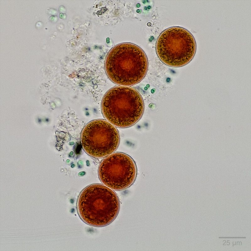
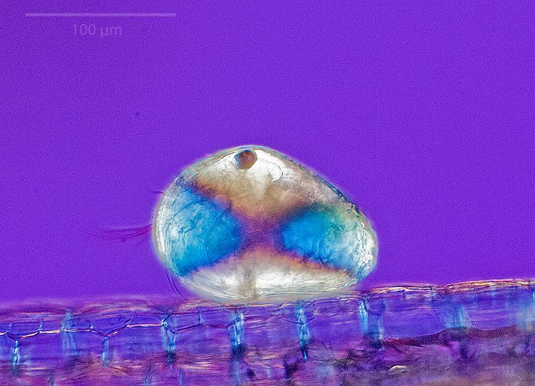
1. Brightfield (with scale bar):
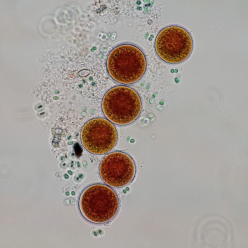
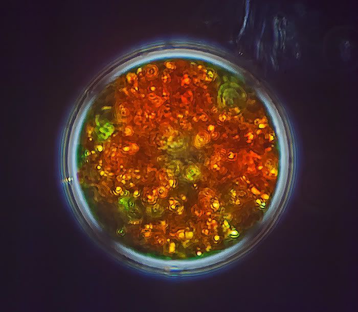
2. Oblique illumination:
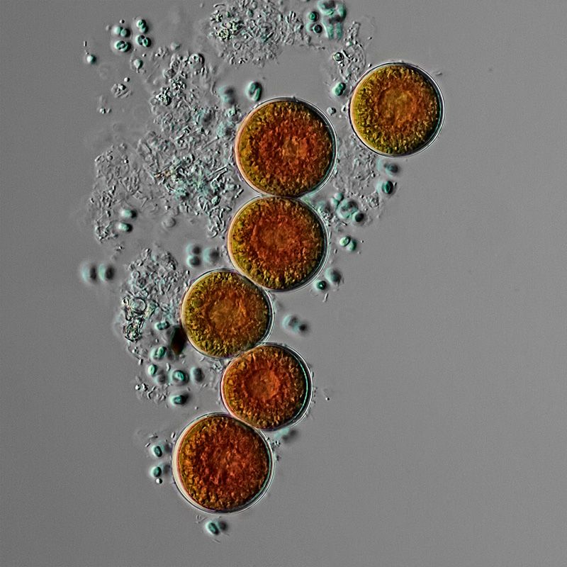
3. DIC:
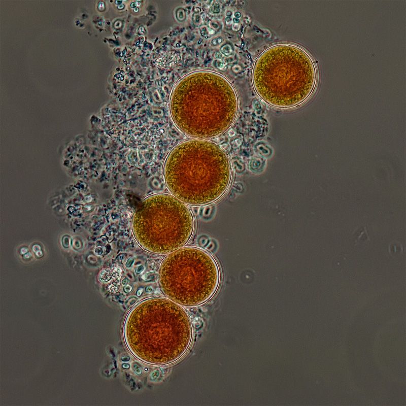
4. Phase contrast:
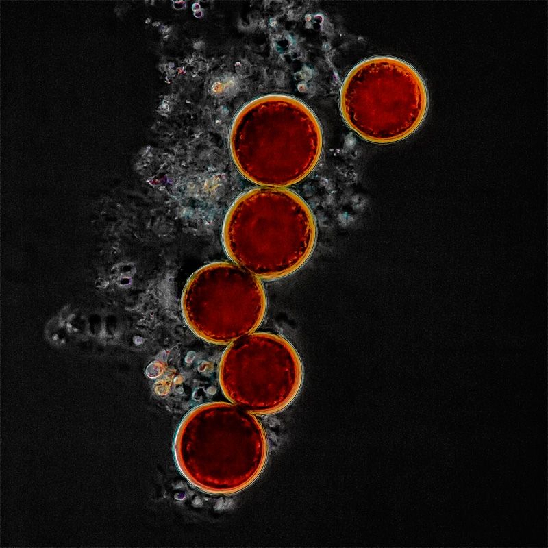
5. Darkfield:
5. Cross-polarized light: [should be 6.]
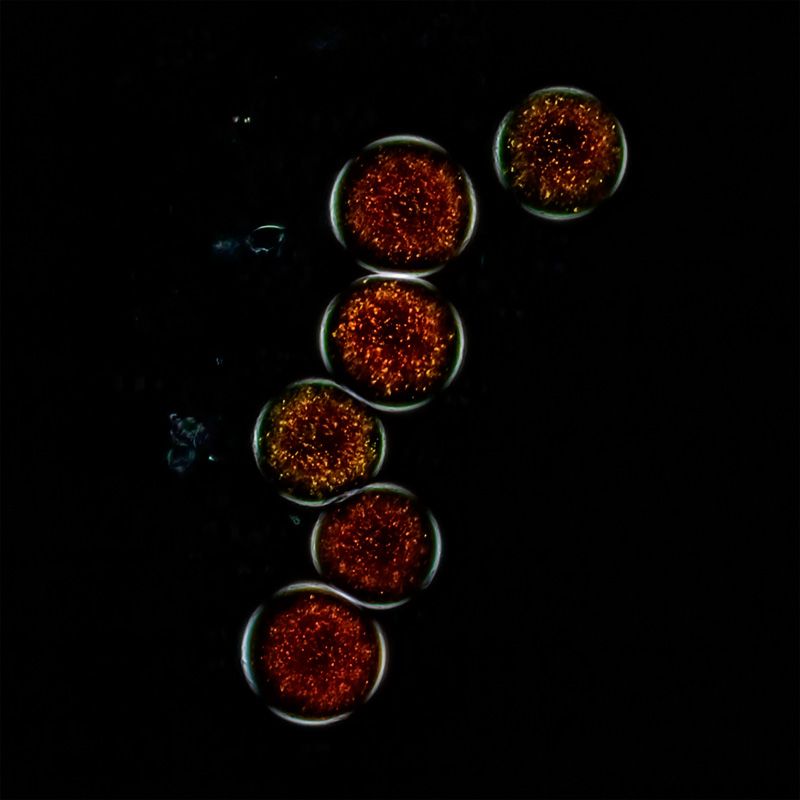
Thanks to Crater Eddie for the water sample containing Haematococcus.

Hi BillT,billbillt wrote:Hi Gekko,
What method do you use to embed your scale bar?..
Thanks!
BillT
Yes.zzffnn wrote:What scope is your DIC on? Is it a Nikon Optiphot?
Focus stacks are not very friendly towards me, so I avoid them whenever possibleBright field is useful for its flat surface resolution - very deep stack of bright field may actually offer more surface details than DIC.
Nikon Fluor 20 Ph3DL.Which 20x 0.75 do you have? I have an old Zeiss Jena 20x 0.65 and love its center resolution.


