I went back at it today, this time with cross-polarized darkfield illumination. However, I kept my objective's iris fully open to maximize NA.
The lineolae on N. oblonga are now very highly contrasted. They are perceptually white/black when optimally illuminated. I have provided a detail view of a deformed stria. I feel like this is worth noting as it shows there is not a continuation of the grating pattern indicating some sort of false image.
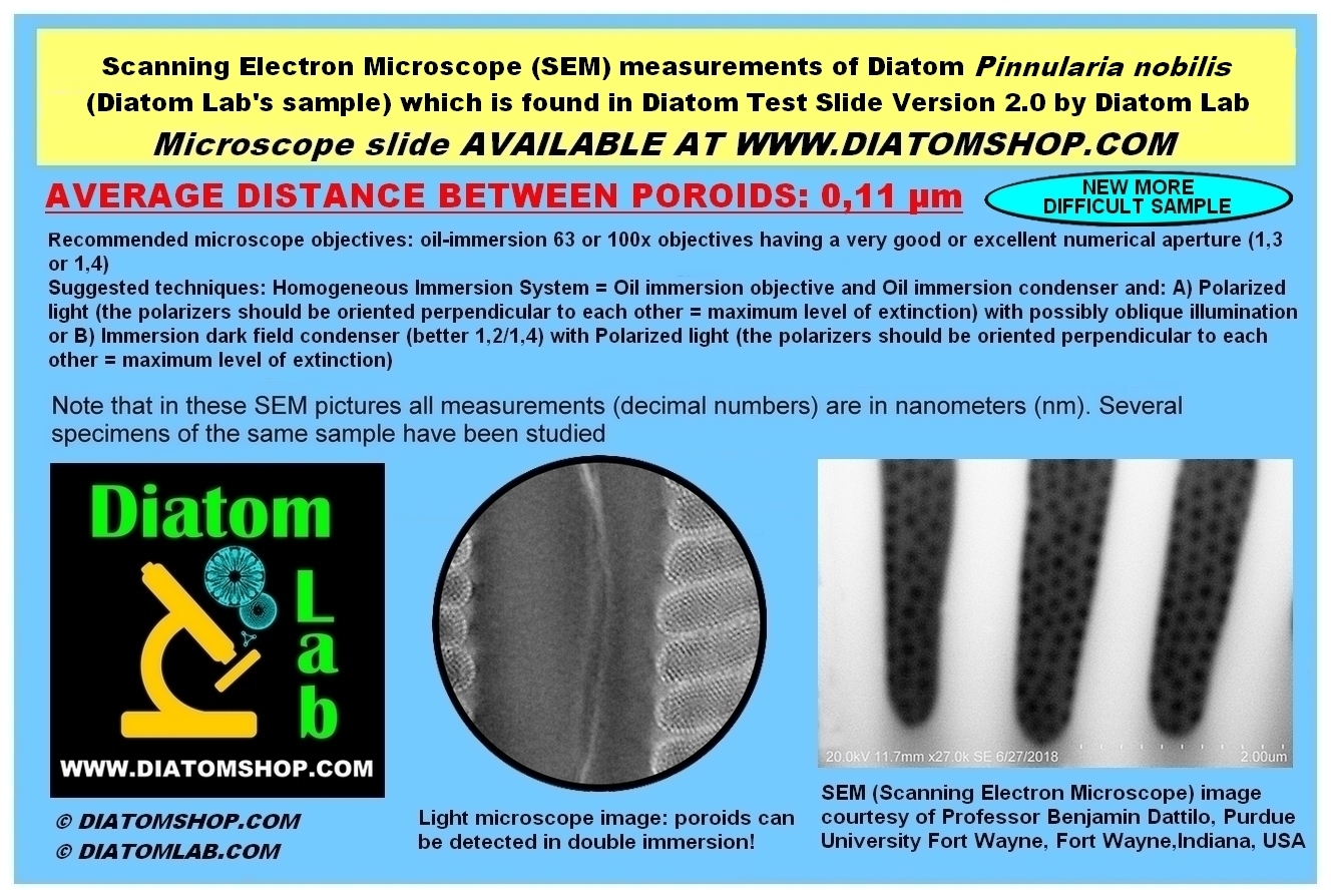
For P. nobilis I attempted a similar approach. The exposures for these subjects are on the order of 10s, so the camera display is extremely noisy. Much of my approach yesterday could be described as trial and error focusing. Today I used the eyepieces more and found some features which I could tell were absolutely definitely real. I saw a constellation of larger pores - two spaced diagonally somewhat far apart and a line of three others nearby. I used this as a reference point when examining the images in post so that, if I saw them, I knew I was actually focused on something. In the zoomed out photo, this constellation is about three quarters over and three quarters down. Refer to the detail image to find it. I know with certainty that those are real features - and I believe the nearby intensity modulations are smaller pores. The checkerboard modulations on the left side striae may be interference artifacts. I have uploaded lossless quality images to the Gdrive:
https://drive.google.com/drive/u/0/fold ... ZCcUS1epOK
I also did some reading up on using high numerical aperture materials to elicit super-resolution. One technique developed in that last couple decades is using microspheres of high index material in contact with the subject. These couple evanescent EM waves which would normally only exist near the surface of the subject and cause them to propagate as light. Most papers I saw using this method stated a maximum resolution of down to 100-80nm which would agree well with diatom lab's stated resolutions. I wonder if their proprietary mountant is an adhesive (NOA170?) mixed with colloidal high RI particles? Or do you even need them if you're already using a high NA mountant? I don't imagine they're inclined to be forthcoming about their trade secret


- noblonga-dfpol-sq.jpg (112.83 KiB) Viewed 6550 times

- noblonga-dfpol-sq-detail.jpg (155.08 KiB) Viewed 6550 times

- pinobilis-dfpol-sq.jpg (160.45 KiB) Viewed 6550 times

- pinobilis-dfpol-sq-detail.jpg (221.4 KiB) Viewed 6550 times


