
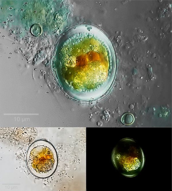
[Edit] I forgot to say that the first image is a focus stack of 15 images taken at 1 µm focus steps and stacked using CombineZP.

How are you getting 1µm focus steps?[Edit] I forgot to say that the first image is a focus stack of 15 images taken at 1 µm focus steps and stacked using CombineZP.
Thank you!Mintaka wrote:A very nice picture indeed. The reddish chloroplast makes me want to say Haematococcus. Also, thanks for drawing attention to "CombineZP" ... I'll be sure to check it out.
Thank you. I've never been happy with the results I get from my 100x objective, so you're saying that it gave a good image makes me happier75RR wrote:At 10µm that is very impressive.
I relied on the calibration marks on the fine focus knob; I guess I should've said "approximately 1 µm"75RR wrote:How are you getting 1µm focus steps?
Many thanks, Rodney. I use a µ4/3 ("mirrorless") Olympus E-P1 camera body and a 2x projection lens in the phototube.Rodney wrote:Very nice clean images, what type of camera are you using?
Got me wondering what the fine focus graduations represented on my Zeiss RA.I relied on the calibration marks on the fine focus knob; I guess I should've said "approximately 1 µm" :)
I sometimes do that, but for objects with significant depth, I don't know how that would affect the final appearance.75RR wrote:P.S. I do not focus stack by using the graduations on the focusing dial but do it visually - in small steps looking to "carpet" the sample with small focused areas which hopefully the stacking software will pick up.
Thank you very much for the very encouraging comments! I think you and Mintaka are probably correct, but I still don't see the resemblance to the Haematococcus that I've seen in the past or to the images I posted earlier (the link is in my reply to Mintaka above), or to those posted by CE. But then I am a very poor judge of those crittersJimT wrote:First of, beautiful images! Well worth the effort with the OI obj.
Second I agree with Mintaka, looks like images of Haematococcus on the web and is the right size. Surprised you couldn't see the flagella in the DIC image.
Others (including you) have posted images in the past that look like these. I will have to get a bird bath
You are correct. But the resting stage, as far as I've seen, is always perfectly round in shape. The image above is rather oval.Crater Eddie wrote:In the dormant, or "encysted" stage, haematococcus do not have flagella, or at least I have never seen one at this stage.


