
Here I present to you my findings from the salt water aquarium.
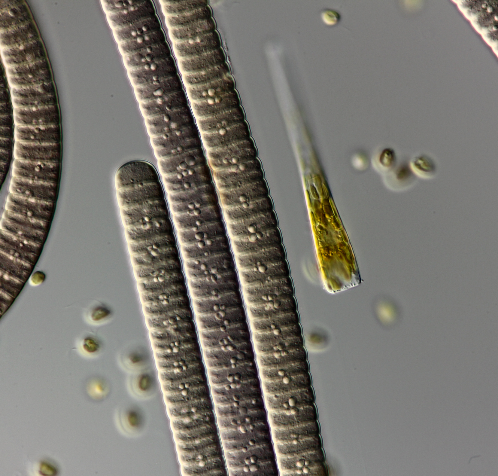
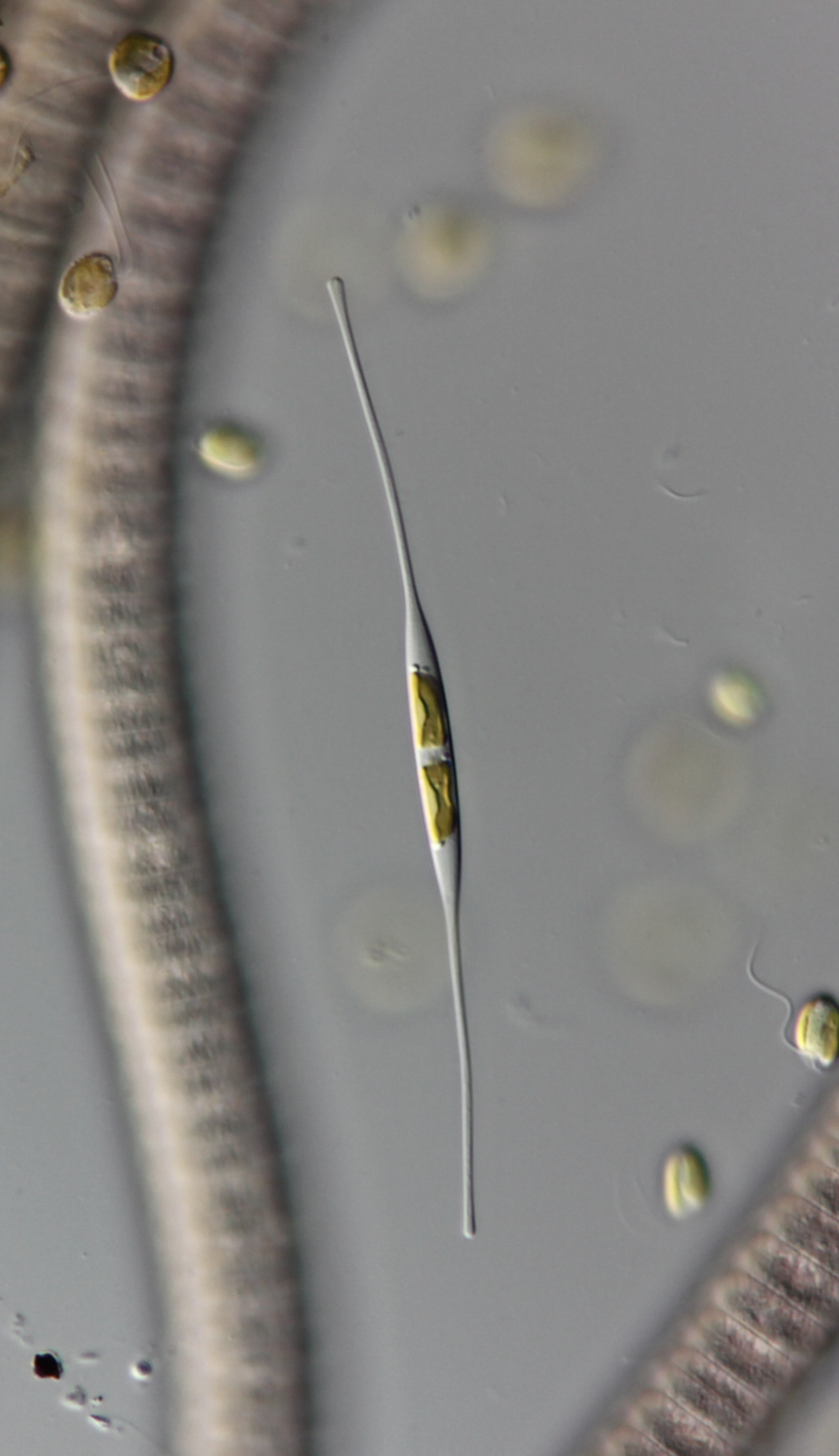
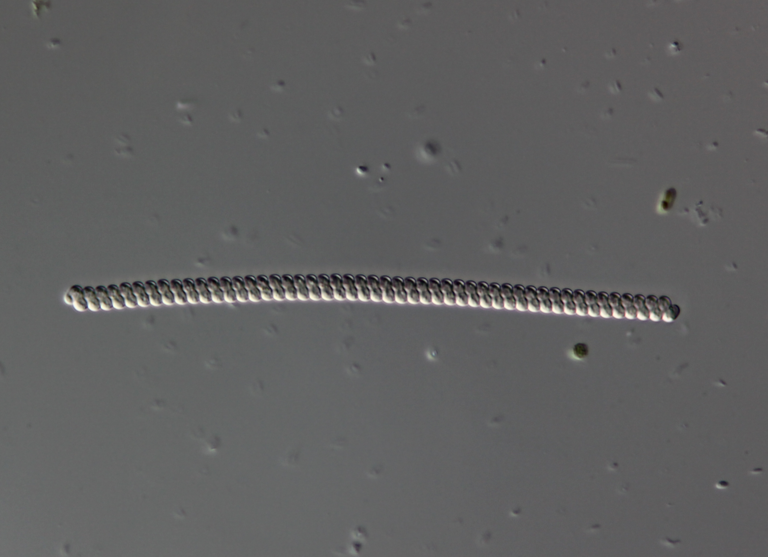
The bacteria, algae and diatoms
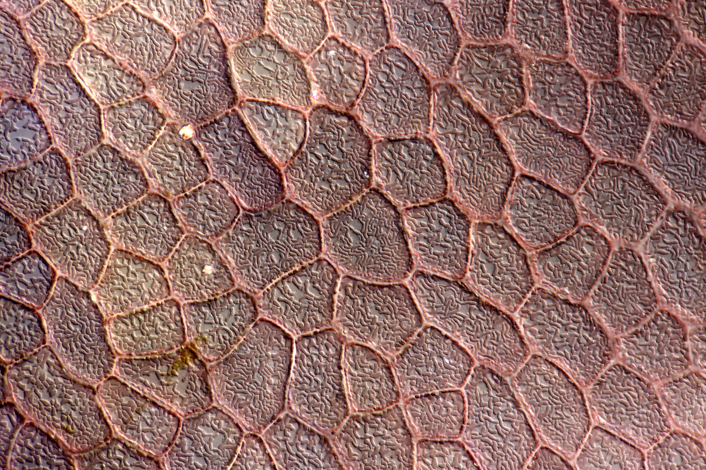
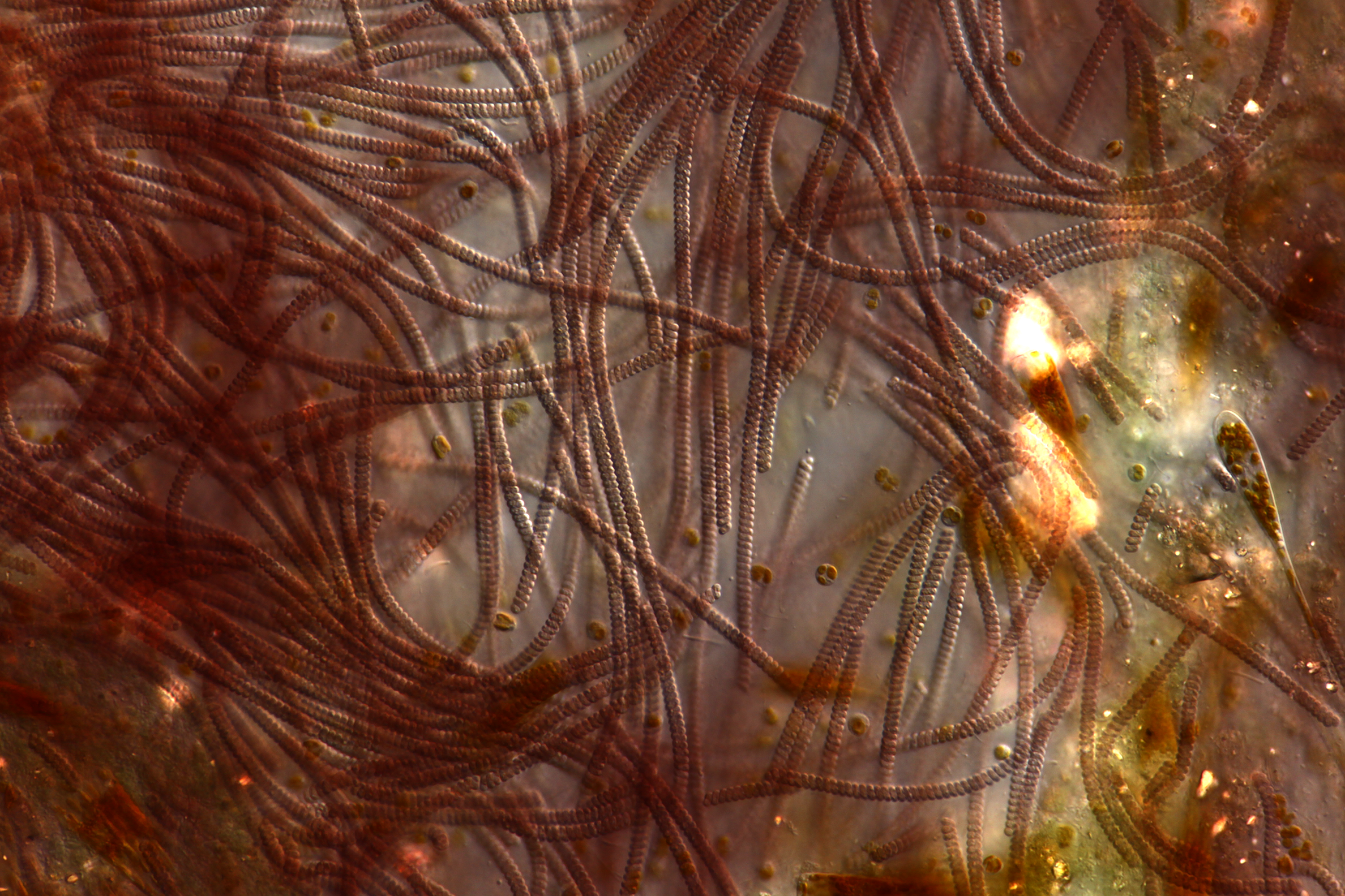
(Red algae)
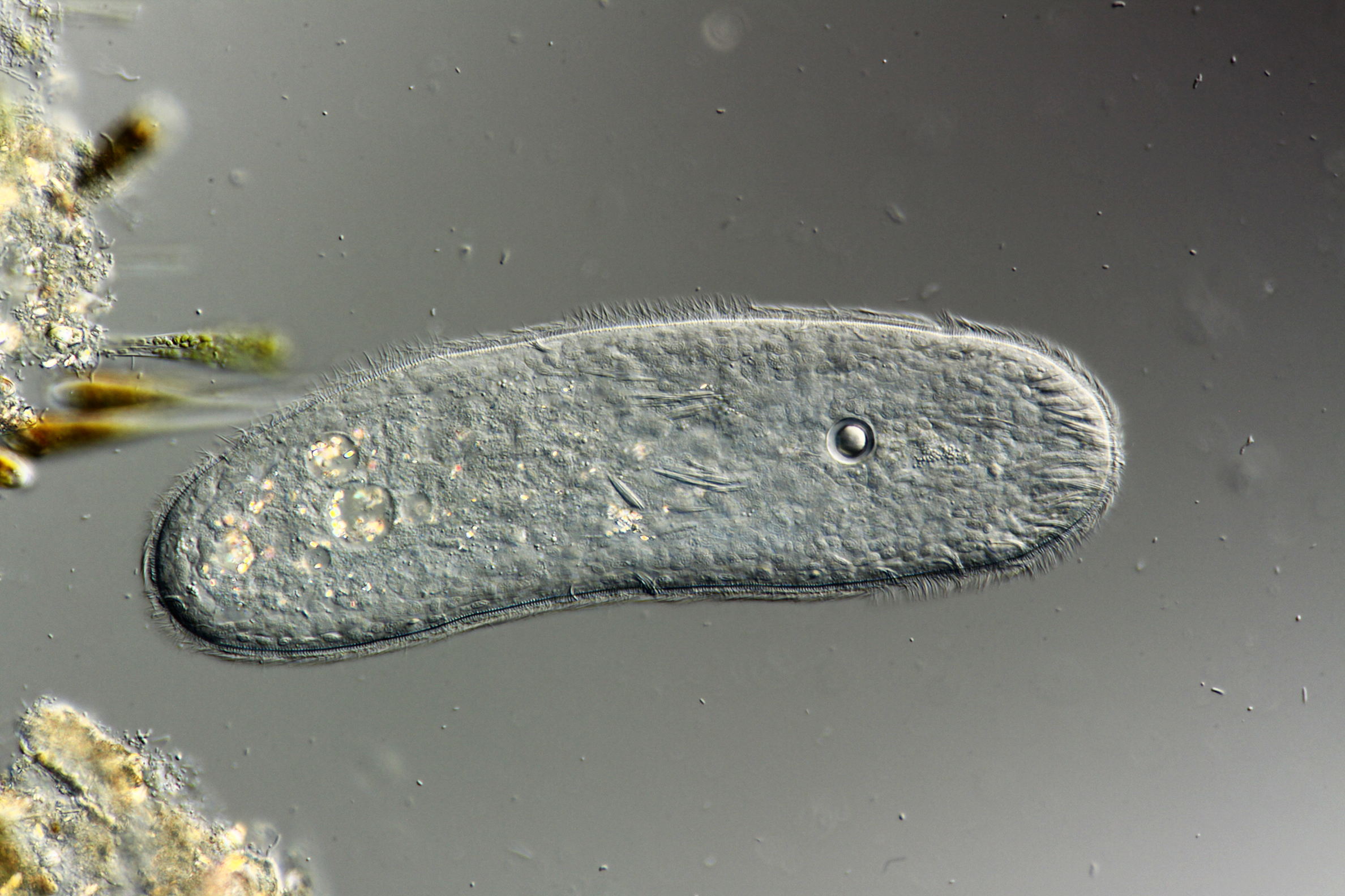
The protists
(Not at all sure what this is)
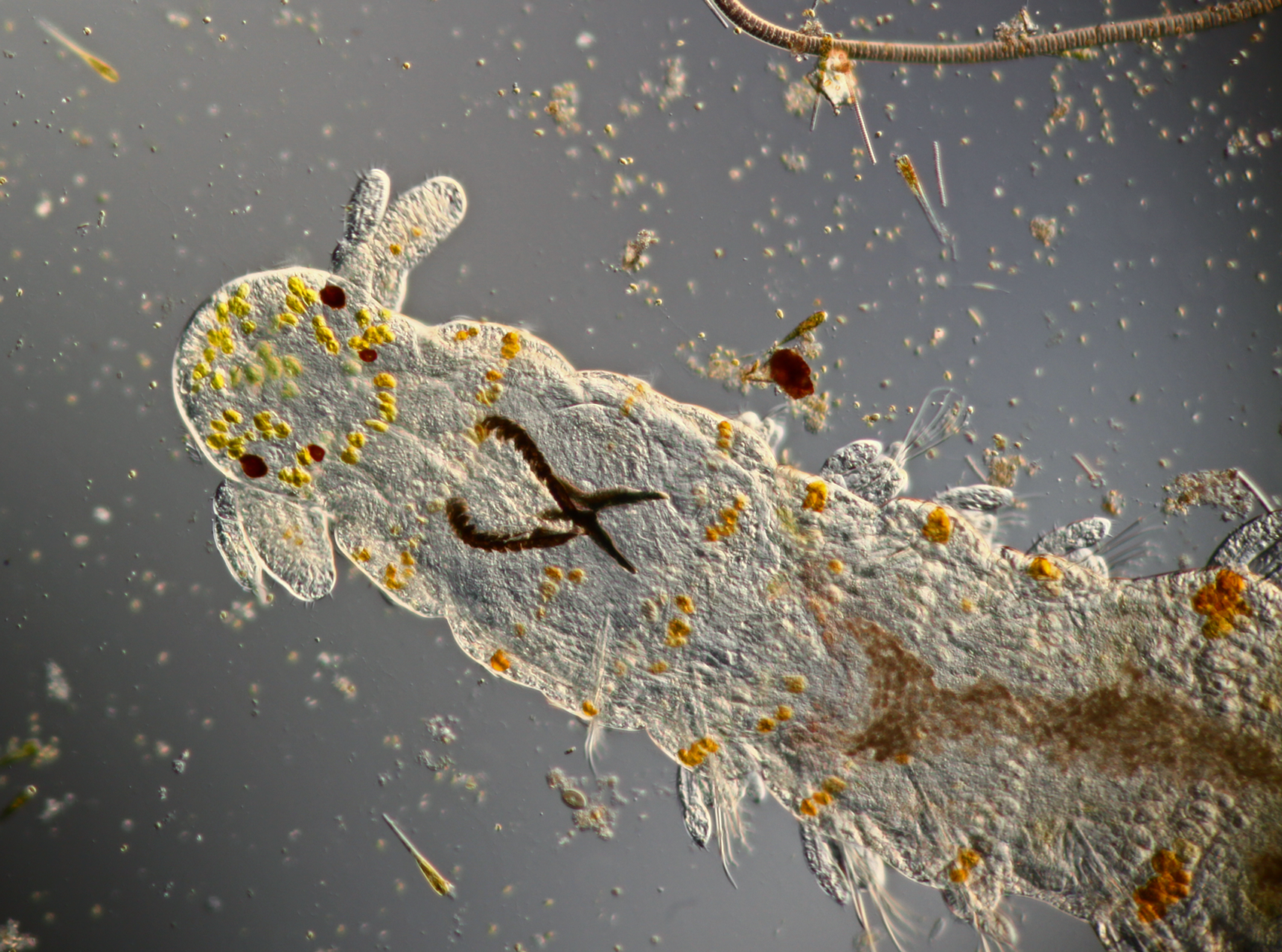
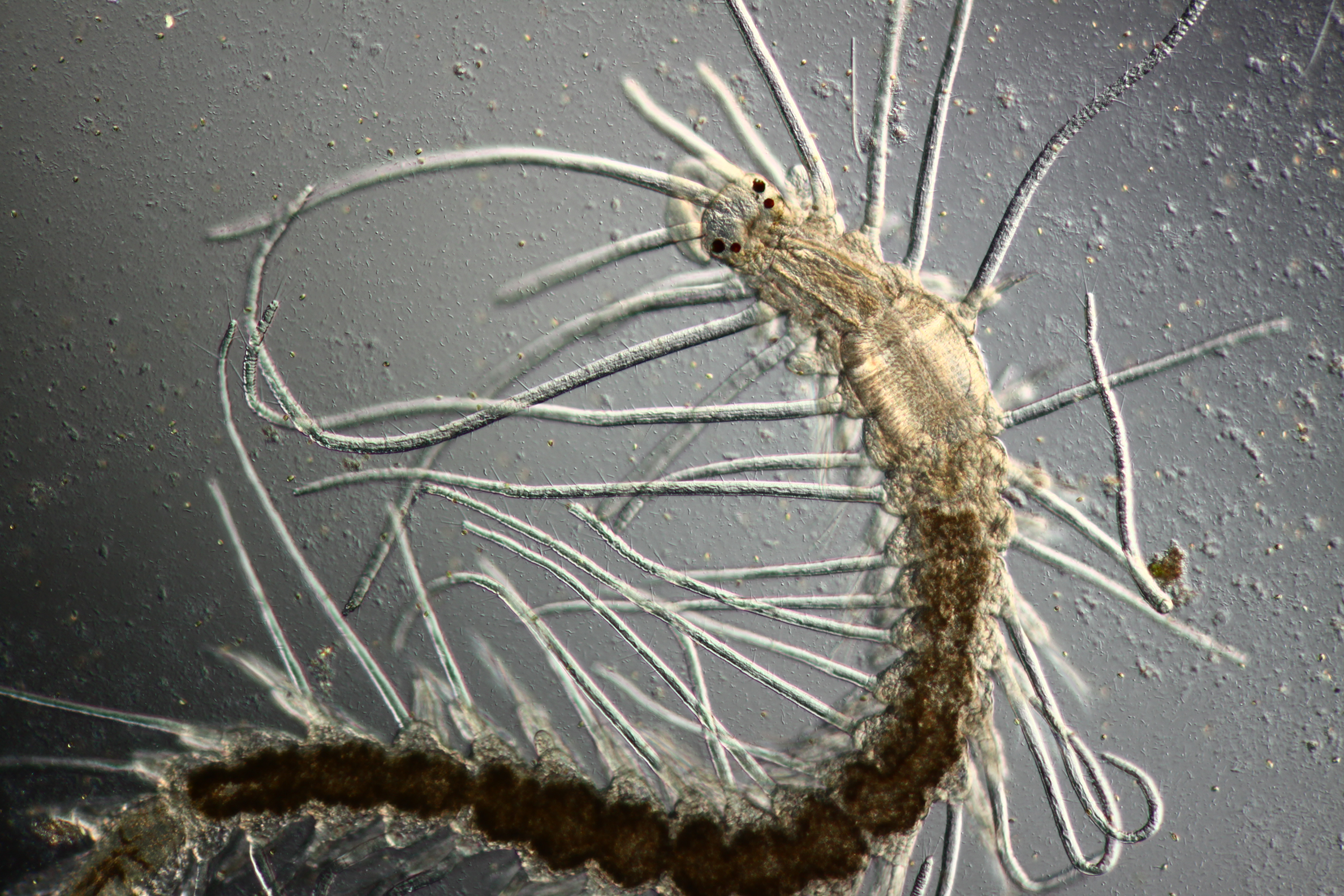
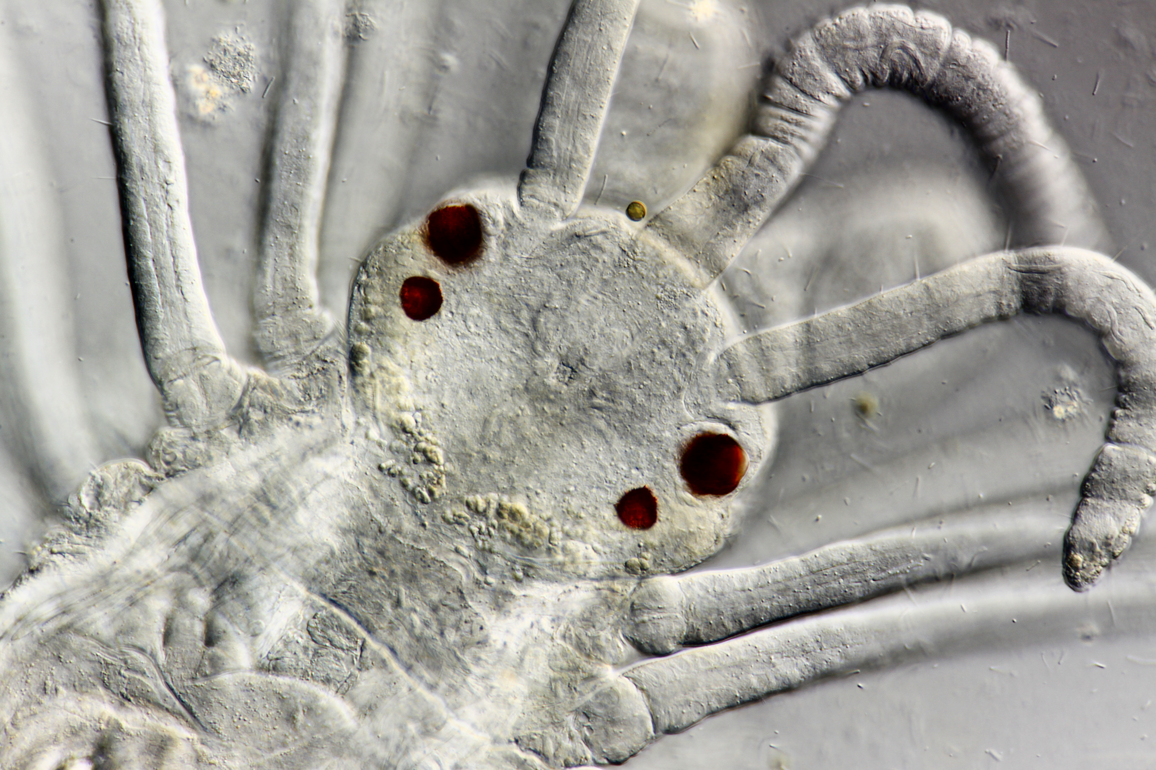
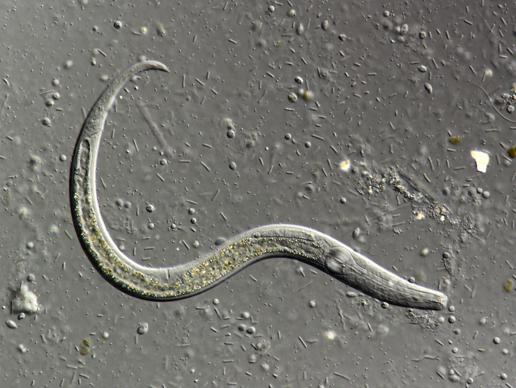
The worms
Best regards,
Wes
Usual equipment, but I did get a DIC prism that works well with my ancient 25x planapo and used that for some of the images.
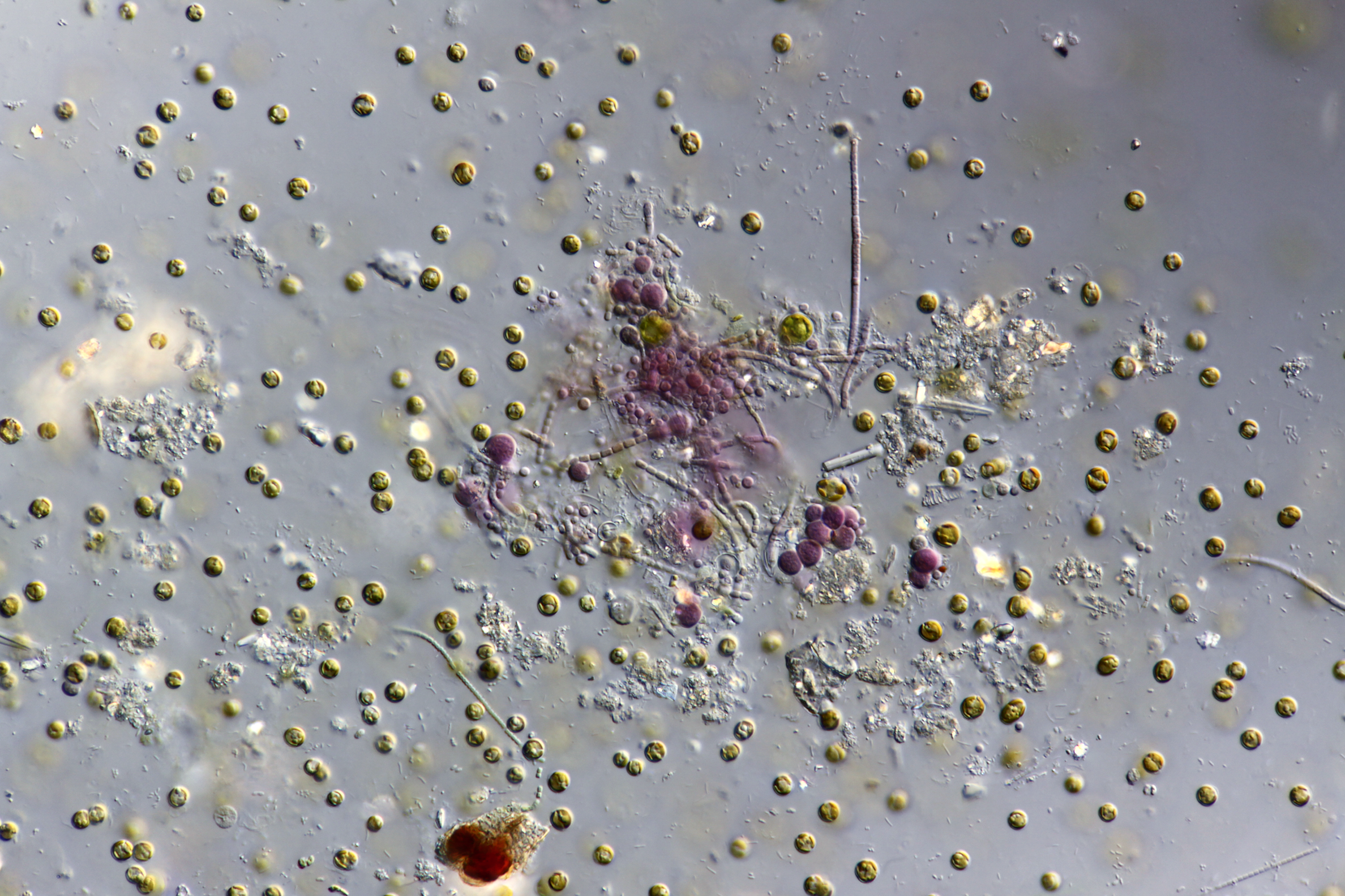
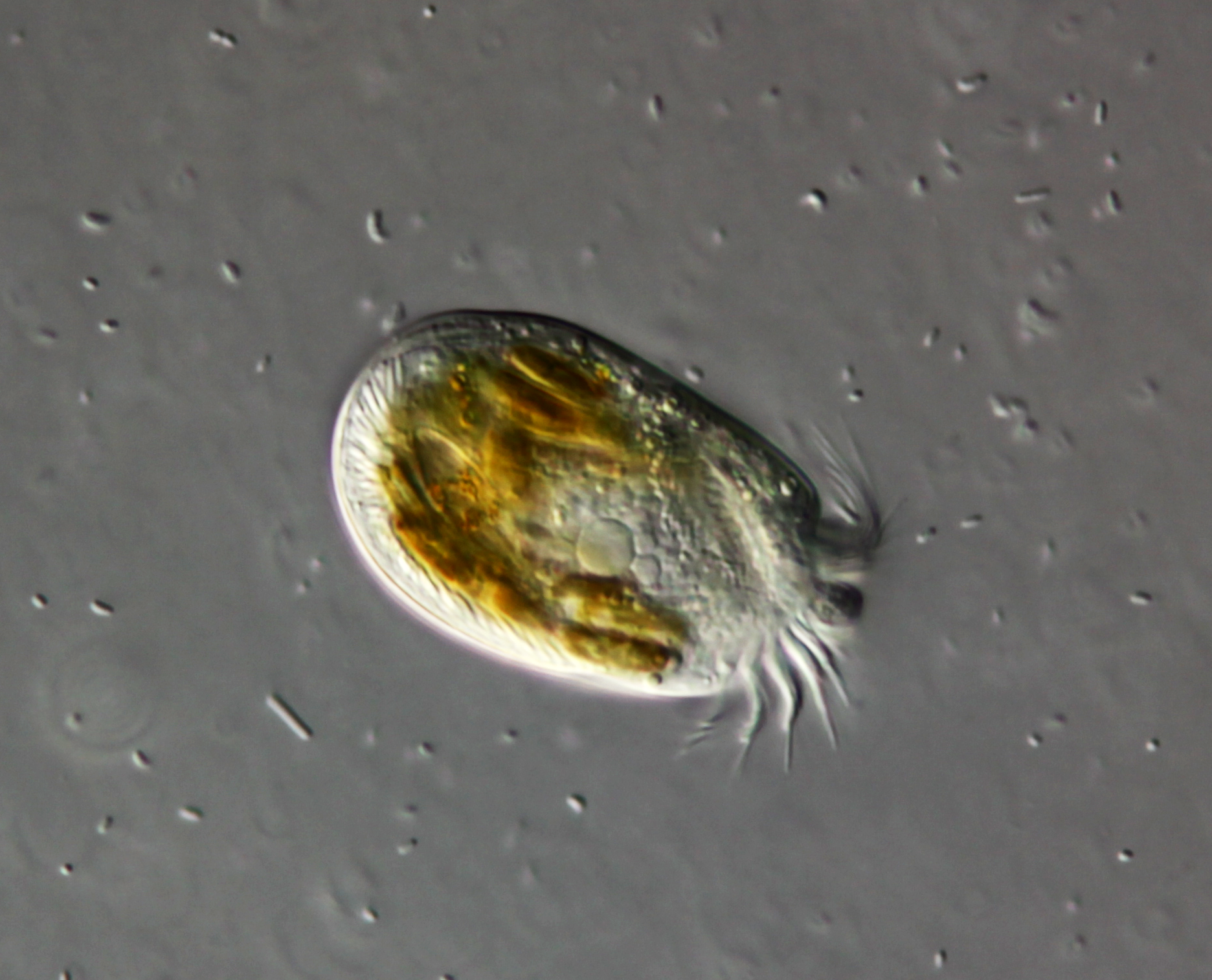
Honestly I have no idea what the oval ciliate is, frankly I'm not sure if its even a protists, could be the larval stage of something. The round object in its right side could be a Müller vesicle or a statolith. I've been entertaining the idea of isolating such unusual things under a stereomicroscope to amplify and sequence its ribosomal genes for a more reliable ID... maybe some day I will give this a try. As for the purple things... well its purple and its bacteria but beyond that I cannot speculate much (maybe some sort of rhodopsin-containing photosynthetic bacteria). I am really unfamiliar with marine life (which makes it way more exciting).

Interesting! I had the idea Müller vesicles were only present on Loxodes. The oval microorganism looks a lot like a ciliate to me with all those distinctive cilia around the cell membrane and those vacuoles. Maybe someone will chime in to clarify this.Wes wrote: ↑Tue Sep 27, 2022 8:23 pmThank you all for the comments and interest.
Usual equipment, but I did get a DIC prism that works well with my ancient 25x planapo and used that for some of the images.
Honestly I have no idea what the oval ciliate is, frankly I'm not sure if its even a protists, could be the larval stage of something. The round object in its right side could be a Müller vesicle or a statolith. I've been entertaining the idea of isolating such unusual things under a stereomicroscope to amplify and sequence its ribosomal genes for a more reliable ID... maybe some day I will give this a try. As for the purple things... well its purple and its bacteria but beyond that I cannot speculate much (maybe some sort of rhodopsin-containing photosynthetic bacteria). I am really unfamiliar with marine life (which makes it way more exciting).
On another forum it was pointed out to me that the round structure is in fact a statolith likely belonging to a member of Turbellaria or Acoelomorpha, not a protist at all.Javier wrote: ↑Tue Sep 27, 2022 9:40 pmInteresting! I had the idea Müller vesicles were only present on Loxodes. The oval microorganism looks a lot like a ciliate to me with all those distinctive cilia around the cell membrane and those vacuoles. Maybe someone will chime in to clarify this.Wes wrote: ↑Tue Sep 27, 2022 8:23 pmThank you all for the comments and interest.
Usual equipment, but I did get a DIC prism that works well with my ancient 25x planapo and used that for some of the images.
Honestly I have no idea what the oval ciliate is, frankly I'm not sure if its even a protists, could be the larval stage of something. The round object in its right side could be a Müller vesicle or a statolith. I've been entertaining the idea of isolating such unusual things under a stereomicroscope to amplify and sequence its ribosomal genes for a more reliable ID... maybe some day I will give this a try. As for the purple things... well its purple and its bacteria but beyond that I cannot speculate much (maybe some sort of rhodopsin-containing photosynthetic bacteria). I am really unfamiliar with marine life (which makes it way more exciting).
Hi Kurt, thank you for the comment and yes I sent the images to the store owner who was fascinated by the invisible biology of their aquarium.
I fine lens indeed, what sort of trans DIC combination have you come up with?
Wow, flat worm! And just when I thought I knew a little about microorganisms...Bruce Taylor wrote: ↑Wed Sep 28, 2022 11:36 pmMarvellous images!
The first ciliate is in the family Strombidiidae (a very abundant group, in salt water).
The pigmented fellow is a species of Pseudokeronopsis (e.g. P. rubra)
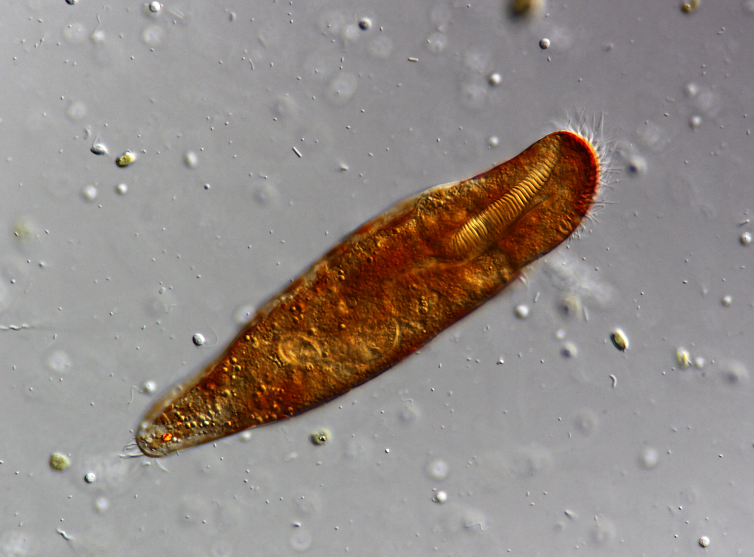
As you already know, the next ciliated creature is not a ciliate at all, but an acoel (acoelomorph) flatworm, equipped with a statocyst
Just this week I received a condenser and slider of the first generation. The parts are a little beaten up but the results are great so far, despite not using the correct objectives.I fine lens indeed, what sort of trans DIC combination have you come up with?
Yes, acoelomorphs certainly can look like ciliates. They even have digestive vacuoles, instead of a gut. Very odd creatures!
I was hoping you would pop by and drop some IDs, thanks a lot!Bruce Taylor wrote: ↑Wed Sep 28, 2022 11:36 pmMarvellous images!
The first ciliate is in the family Strombidiidae (a very abundant group, in salt water).
The pigmented fellow is a species of Pseudokeronopsis (e.g. P. rubra)
As you already know, the next ciliated creature is not a ciliate at all, but an acoel (acoelomorph) flatworm, equipped with a statocyst
I didGreg Howald wrote: ↑Thu Sep 29, 2022 12:48 amI hope you will share them with the guy who gave you the samples.
Thank you!Bruce Taylor wrote: ↑Thu Sep 29, 2022 8:15 pmYes, acoelomorphs certainly can look like ciliates. They even have digestive vacuoles, instead of a gut. Very odd creatures!
In this case, the statocyst--that single round organ on the centerline of the cell, in the anterior--is a pretty strong clue. Also, those shaggy spindle-shaped organs on its body do not resemble ciliate extrusomes or other organelles. I don't know much about worms, but I think these could be "rhabdoids" (whatever those might be). The marine habitat is typical for acoelomorphs, as far as I know.

