Here you can post pictures and videos to show others.
-
hkv
- Posts: 1012
- Joined: Sun Nov 02, 2014 8:57 pm
- Location: Sweden
-
Contact:
#1
Post
by hkv » Sun Apr 01, 2018 9:19 am
Lucky to have found Micrasterias in my samples, which are very beautiful (and big). Could not fit the entire subject with the 40X, so the full body shots are taken with a 20X objective. I just acquired a new oil darkfield condenser so this is the first image with the new toy.
20X, Polarized light:
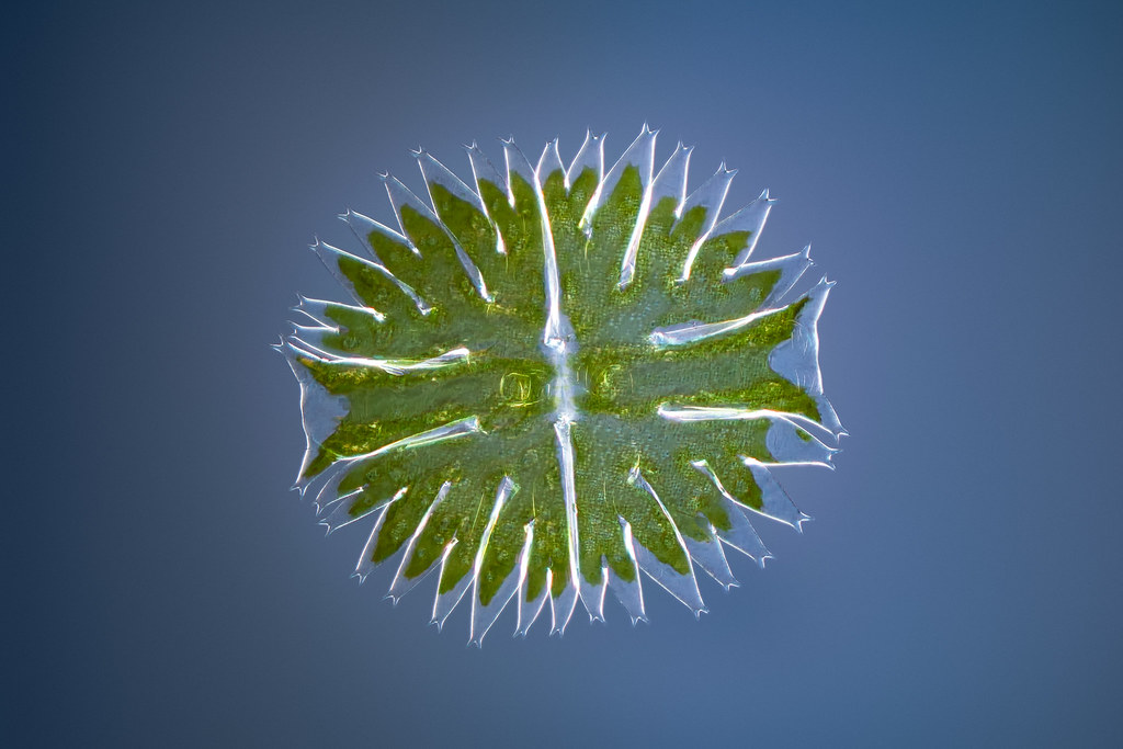 Green algae - Micrasterias
 by
Håkan Kvarnström, on Flickr
20X, DIC:
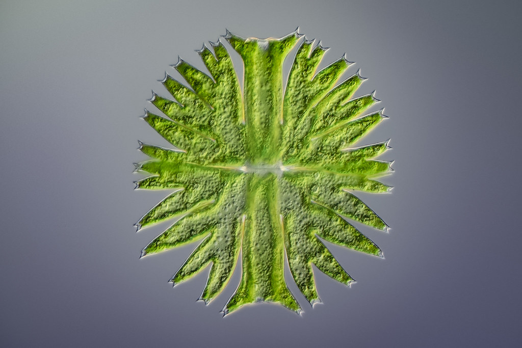 Green algae - Micrasterias
 by
Håkan Kvarnström, on Flickr
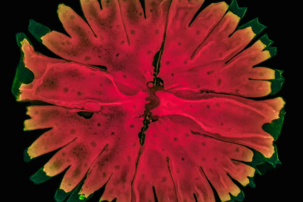
20X, Fluorescent light. Showing the chloroplasts in red:
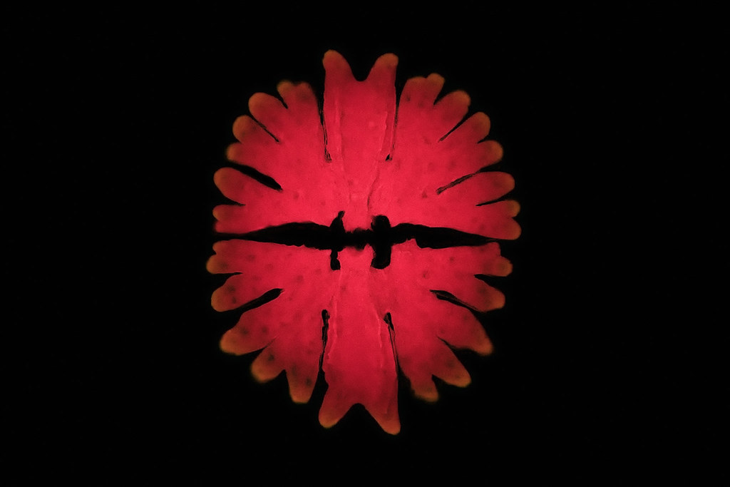 Green algae - Micrasterias
 by
Håkan Kvarnström, on Flickr
20X, Darkfield:
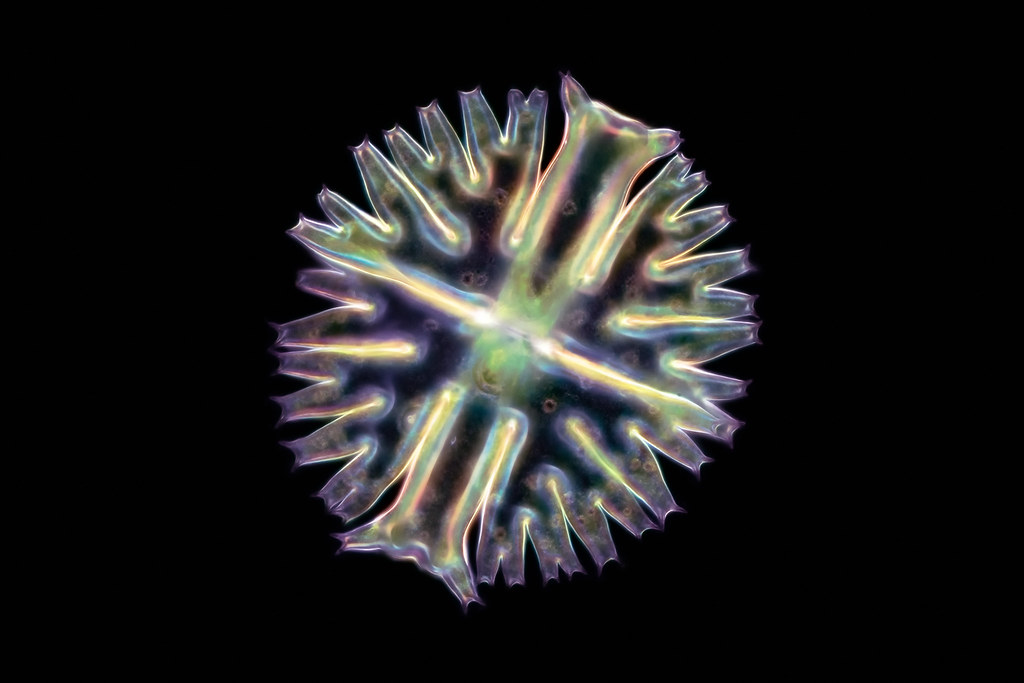 Green algae - Micrasterias
 by
Håkan Kvarnström, on Flickr
40X, Fluorescent light:
 Green algae - Micrasterias
 by
Håkan Kvarnström, on Flickr
-
Hobbyst46
- Posts: 4296
- Joined: Mon Aug 21, 2017 9:02 pm
#2
Post
by Hobbyst46 » Sun Apr 01, 2018 9:30 am
@hkv
Very nice and impressive work.
I wonder which wavelength you used to excite the red fluorescence, and which setup (lamp, filter cubes etc). Are these on the Olympus BX51? I am asking since there are several possible wavelengths, and several chlorophyll and related molecules.
-
75RR
- Posts: 8207
- Joined: Sun Oct 12, 2014 2:34 am
- Location: Estepona, Spain
#3
Post
by 75RR » Sun Apr 01, 2018 10:54 am
Great catch + great images
Have not seen one for ages
Have you thought about using the 40x and combining the partial images via stitching?
Note: stitching software requires a large image overlap
Zeiss Standard WL (somewhat fashion challenged) & Wild M8
Olympus E-P2 (Micro Four Thirds Camera)
-
hkv
- Posts: 1012
- Joined: Sun Nov 02, 2014 8:57 pm
- Location: Sweden
-
Contact:
#4
Post
by hkv » Fri Apr 06, 2018 3:07 pm
Hobbyst46 wrote:@hkv
Very nice and impressive work.
I wonder which wavelength you used to excite the red fluorescence, and which setup (lamp, filter cubes etc). Are these on the Olympus BX51? I am asking since there are several possible wavelengths, and several chlorophyll and related molecules.
Yes, Olympus BX51. I used a mercury lamp (
http://www.excelitas.com/Pages/Product/ ... xacte.aspx)
Used a filter cube having the following specifications:
Excitation filter: 440-440 nm
Dichroic mirror: 455 nm
Emission: 475 nm
-
hkv
- Posts: 1012
- Joined: Sun Nov 02, 2014 8:57 pm
- Location: Sweden
-
Contact:
#5
Post
by hkv » Fri Apr 06, 2018 3:10 pm
75RR wrote:Great catch + great images
Have not seen one for ages
Have you thought about using the 40x and combining the partial images via stitching?
Note: stitching software requires a large image overlap
Thank 75RR. I have several stitches shot, but I have not had the time to post process them yet. Also, I swapped out the 2.5X photo eyepiece to a 2X photo eyepiece and JUST managed to get small examples of the Micrasterias in one shot! It was pixel close, but it worked. Will post these later also when I find time to spend on my hobbies...
-
Hobbyst46
- Posts: 4296
- Joined: Mon Aug 21, 2017 9:02 pm
#6
Post
by Hobbyst46 » Fri Apr 06, 2018 3:58 pm
hkv wrote:
Excitation filter: 440-440 nm
Dichroic mirror: 455 nm
Emission: 475 nm
The 440 is quite inside the excitation range for chlorophylls and pheophytins, but other molecules will also be excited.. The dichroic mirror and emission filters are also not specific for chlorophylls, but allow you to observe the fluoresence of other molecules in the green-yellow light range, as is nicely visible in the 40x fluorescence image. To isolate just the chlorophylls one would need a dichroic of about 550 and a 600nm emission filter (just a side note. the images are superb as they are). I am surprised that the yellow and green are not visible in the 20x objective image.
There are vitamin E - related pigments in some plants that fluoresce in the 500-600nm - I wonder if the tiny alga contains them...anyway I note that the chlorophylls dominate the emission.
-
hkv
- Posts: 1012
- Joined: Sun Nov 02, 2014 8:57 pm
- Location: Sweden
-
Contact:
#7
Post
by hkv » Fri Apr 06, 2018 4:26 pm
Hobbyst46 wrote:hkv wrote:
Excitation filter: 440-440 nm
Dichroic mirror: 455 nm
Emission: 475 nm
The 440 is quite inside the excitation range for chlorophylls and pheophytins, but other molecules will also be excited.. The dichroic mirror and emission filters are also not specific for chlorophylls, but allow you to observe the fluoresence of other molecules in the green-yellow light range, as is nicely visible in the 40x fluorescence image. To isolate just the chlorophylls one would need a dichroic of about 550 and a 600nm emission filter (just a side note. the images are superb as they are). I am surprised that the yellow and green are not visible in the 20x objective image.
There are vitamin E - related pigments in some plants that fluoresce in the 500-600nm - I wonder if the tiny alga contains them...anyway I note that the chlorophylls dominate the emission.
Yes, I was not really trying to excite only the chlorophyll. I think it is great if everything that can lit up and becomes visible. When it comes to the green excitation, you can see in the 40X that the green rim is very noisy. The camera sensor barely registered the signal and most was noise. I enhanced that in post as much as possible. In the 20X I rather supressed the green in post because it was not visible to the naked eye. Only if I pushed shadows to max you could see traces of the green.
Last edited by
hkv on Fri Apr 06, 2018 5:04 pm, edited 2 times in total.
-
billbillt
- Posts: 2895
- Joined: Mon Nov 10, 2014 10:01 pm
#8
Post
by billbillt » Fri Apr 06, 2018 5:02 pm
Very, very good... Thanks for posting...
BillT
-
MicroBob
- Posts: 3154
- Joined: Sun Dec 25, 2016 9:11 am
- Location: Northern Germany
#10
Post
by MicroBob » Fri Apr 06, 2018 7:09 pm
Hi Håkan,
great images again!
The darkfield image is not what I would have expected - your micrasterias looks more like a diatom!
Here in Hamburg they have a big collection of these algae at the botanical instute of the university. If we ask friendly, they breed a couple of species for our microscopy group. These algae were collected around the world by a woman in the 1960s. Since then they are kept at the University. They multiply very slowly but from time to time a new start has to be made. I find it incredible that they have managed to keep them alive for half a century!
Probably there is a "special treatment" for students who accidentally destroy one of the cultures!

I think this type of algae is mostly found in moor water?
Bob
-
vasselle
- Posts: 2763
- Joined: Sat Oct 25, 2014 5:32 pm
- Location: France
#11
Post
by vasselle » Tue Apr 10, 2018 6:43 pm
Bonjour
Superbes images
Cordialement seb
Microscope Leitz Laborlux k
Boitier EOS 1200D + EOS 1100D
-
SunshineLW
- Posts: 107
- Joined: Sat Mar 03, 2018 5:30 pm
- Location: College Station, TX
-
Contact:
#12
Post
by SunshineLW » Wed Apr 18, 2018 11:11 am
Absolutely amazing work!
-
desertrat
- Posts: 243
- Joined: Thu Apr 05, 2018 1:06 am
- Location: Idaho
#13
Post
by desertrat » Thu Apr 19, 2018 1:01 am
Those are all amazing images. I think I like the first one best, just from as aesthetic standpoint, even though some of the others show more detail of certain structures.
Rick
A/O 10 Series Microstar
A/O 4 Series Microstar
A/O 4 Series Phasestar
A/O 4 Series Apostar
A/O Cycloptic Stereo
Several old monocular scopes in more or less decrepit but usable condition


