The development cycle of Haematococcus pluvialis is represented as followed (according to ELLIOT, see also footnote 1):
Fig. 1: Palm cell states (a) can divide into either flagellated cells (h) or again palmellacellas, and the resulting cellular cell states (g) grow into" adult "forms (a) and the circulation can begin anew, but if flagellated Cells form, the cycle continues with the formation of young macrozoospores (f), which soon become normal macrozoospores (e). Periodically, these macrozoospores lose their flagella and turn into a palmella-like form (a), although their From this condition, young macrozoospores (f) are formed and the circulation is repeated to infinity.Hematocysts (b) can be formed from macrozoospores (e), although the usual process leads to the formation of palm cells (a). Under certain conditions, microzoospores (c) are formed from red cysts, which soon turn into young palmella-like states (d), which develop into either M acrozoospores (e) or become Palmella states (a). In both cases the circulation starts from here in the usual way. According to ELLIOT.

Below some photos I made in 2016:
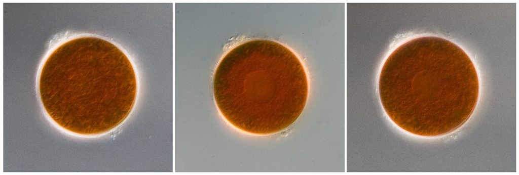
Thick-walled, unspotted and red-colored Aplanospore. The red, cytoplasmic carotenoid astaxanthin overlays existing structures. Depending on the focus, the core could also be seen in the Aplanospore shown here (center photo / right). Maybe the state of development correspont to "(d) - young palmella-like state"?
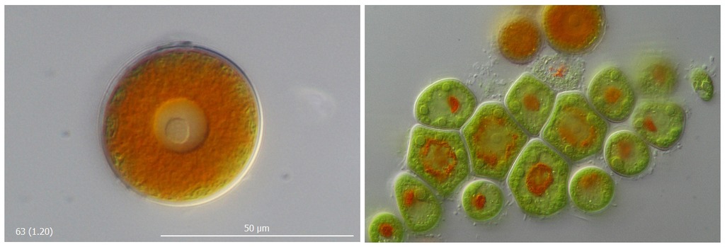
Green, flagellated form, probably "macrozoospores". In addition, intermediate stages with more or less pronounced mucous envelopes.

F Flagellum, G Mucous shell , P Pyrenoid, PF Plasma Precursors, N Nuclei / Core, S Stigma (Eye Patch)

The flagella of hematococcus shall pass through "tubes" through the outer sheath (Huber-Pestalozzi "Volvococcales"). I think they are difficult to observe. Does anybody spotted and documented this tubes? I will try later on my side to find ... I have a pot with red water in the garden
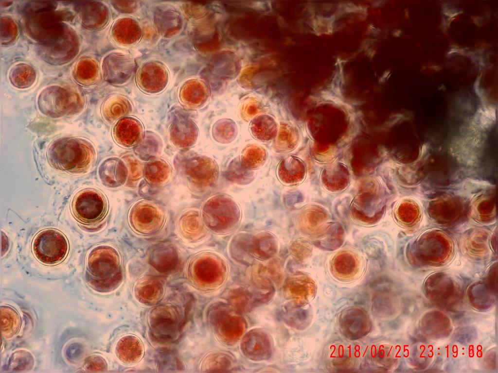
More photos, these show different palmella-like states. The colors vary a lot, ranging from green, yellow, red to brown. I can hardly resist to make too many photos, it is always so existing observing. I try to restrict to show only a few here


Print Media:
(1) Müller, Justus, Kulturversuche mit Haematococcus pluvialis, Mikrokosmos 43, 225 http://www.zobodat.at/pdf/Mikrokosmos_43_0001.pdf
(2) Aumann, G, Haematococcus pluvialis und das Bamberger Blutbecken. Mikrokosmos 43, 99 http://www.zobodat.at/pdf/Mikrokosmos_43_0001.pdf