It's been about three weeks since I put the slide out near the front door with a drop of olive oil, and I put it under the microscope yesterday.
There were particles and fibres although not very many, as Singapore is a city with relatively good pollution control of particles of optical microscopy size range.
I decided to do a quick revision of forensic microscopy on this fibre:
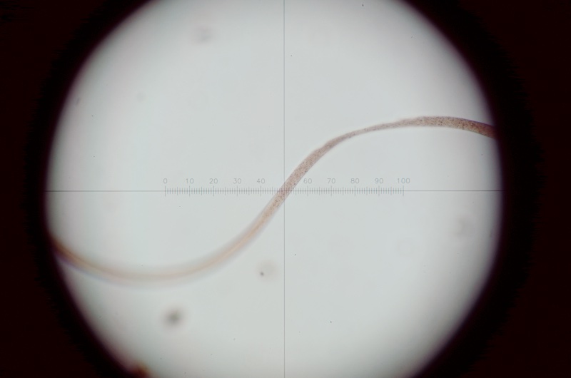
- fibre.JPG (48.98 KiB) Viewed 6748 times
Potentially useful characteristics for fibre identification that can be seen in bright-field include the colour, texture/scale patterns and internal structures (if any) of the fibre. Also the refractive index in the fast and slow directions, which needs to be measured using oils of different refractive index or using dispersion staining, which I'll have to come back to later.
The fibre appears to be of circular cross-section, given its mostly constant width (the thinning out you see above I think is due to damage rather than non-circularity), which is useful for calculating birefringence later. Using the calibrated eyepiece micrometer I measured the fibre diameter/thickness to be 13.5 μm.
By rotating the stage in cross-polarized light I determined that the fibre has straight extinction. This is a sometimes useful clue because some fibres, such as cotton, never go extinct due to their molecular structure.
I also grabbed the chance to use my Berek compensator. I had just taken it apart to make a design modification and reassembled/recalibrated it, so I was keen to put it through its paces:

First, determining the sign of elongation, which helps narrow down the fibre possibilities. The fast ray of my Berek compensator is in the NE/SW direction (opposite to most fixed-wavelength plates) and the fibre's interference colours were subtracted when its length was parallel to that direction, so it's positive elongation (length slow):

Most (but not all) fibres are positive elongation.
Next, I measured the exact retardation value, which is the really fun part of the Berek compensator that involves vernier goniometer readings, checking a table of values from the slide rule era and multiplying the preliminary result by the machine constant that is unique to each compensator:
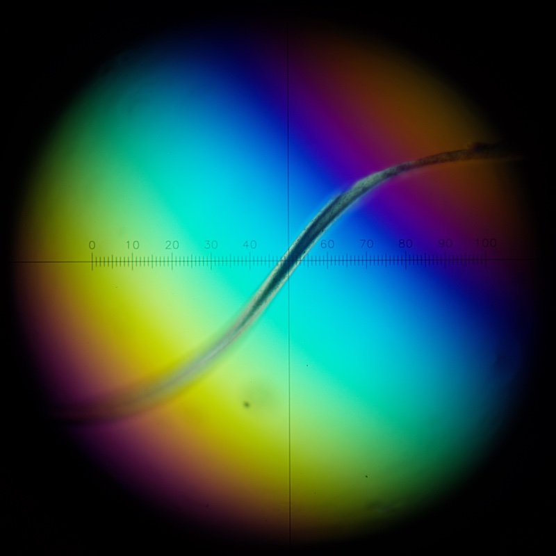
- fibre_retardation.JPG (77.92 KiB) Viewed 6748 times
The calculated retardation value was 706 nm. This, with the measured thickness of the fibre, gives a birefringence value from the Michel-Lévy chart of around 0.048. This can be compared with the reference values of known fibres, which can be animal, plant or synthetic. Natural fibres tend to have higher birefringence than man-made ones, although there are overlaps.
Some of the information I give above came from this book:
Wheeler, B. P. 2021.
Practical Forensic Microscopy: a Laboratory Manual 2nd ed. Wiley.
Another interesting reference:
De Wael, K. 2021.
Microscopy in Forensic Fibre Examinations: a Practical Photo Atlas and Training Tool. Cobalt Blue Coaching.
As mentioned earlier I'm quite interested in the fact that it is possible to identify fibres using a simple optical polarizing microscope, without expensive facilities like Raman spectroscopy or SEM.
I have to move on to a more urgent project for now but I'll try to come back to this and analyze the other stuff on the slide.

