
This post places a focus on what is possible, but does not explain the “how” part. This is something that I plan to include later posts.
Visit the Microscopy Shop!
>>> USA Shop | Germany Shop | UK Shop | Canada Shop <<<
As an Amazon Affiliate, I earn a commission but it does not cost you more.
Stacking
Microscopic images generally have a low depth of field. It is possible to take several images of different depth of fields and to combine them in such a way that the final image is sharp throughout. By carefully turning the fine-focus knob a specified amount, it is possible to section through a complete specimen. Care should be taken, however: If too many parts of a transparent specimen are in focus in the final image, then these parts may cover each other, thus reducing the information content of the final image. Two cell organelles which are located behind each other, both being in focus, will cover up each other, and it is not possible to say which part belongs to what organelle.
Stitching
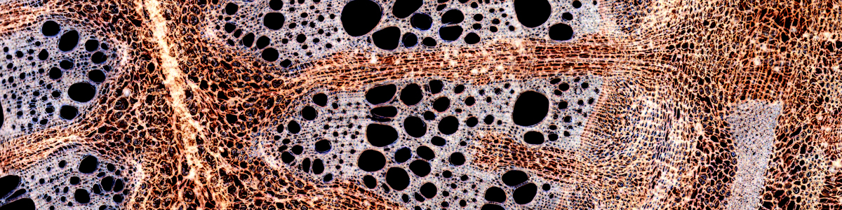
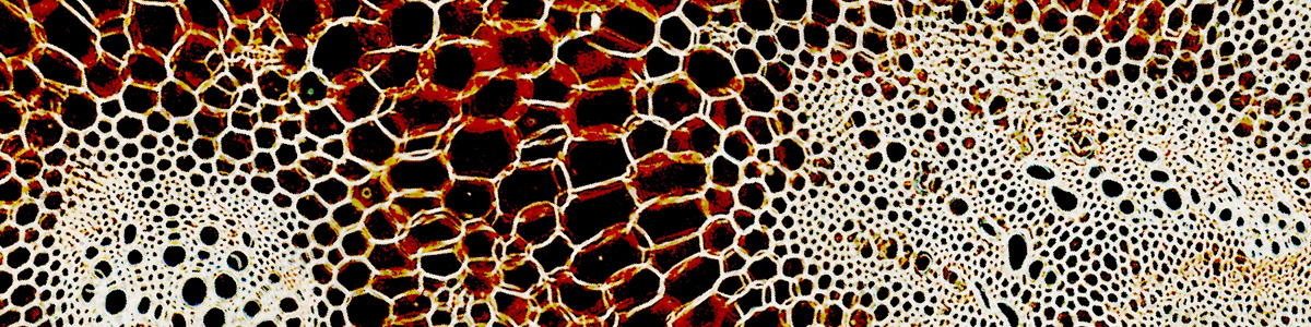
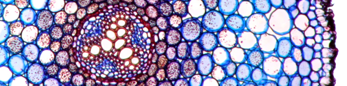
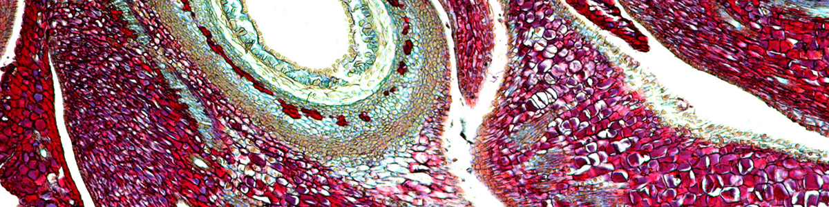
In this method, different overlapping images are assembled together into a larger final picture. While stacking combines the images “vertically”, stitching produces a larger final image by “horizontally” combining them. By stitching, it is possible to overcome the limited field of view. Stitching can be accomplished by using a panorama software. When choosing the software, one should take care that it allows for the combining of images both horizontally and vertically (some only permit for horizontal combination). Microscopic images often do not offer much image complexity. For this reason, the software may have problems assembling the images automatically. It pays off to do a little planning beforehand.
- How large will the final image be? The processing requirements increase significantly with increased image size.
- What camera resolution should be used? The choice of camera resolution has a significant impact on the final image size and required processing power. One should first test, if a high camera resolution is indeed necessary of if it is not simply results in empty magnification. Read: Required camera resolution for photography through the microscope
- How much image overlap should be used? More overlap may make it easier for the software to automatically assemble the pictures, but at the same time more pictures are needed to cover the whole specimen (which again increases work time).
All of the images of the category Virtual Microscope were stitched together.
Background clean up
The optical surfaces (especially the lighting system and condenser optics) are rarely completely free of dust. These disturbances will be present in the image, whether or not a specimen slide is present. It is now possibly to mathematically subtract these disturbances from the image. A picture with and without a specimen has to be taken at the same magnification and using the same exposure time. The empty image (without specimen) is then subtracted from the image containing the specimen.
Alternatively, it is possible to clean the background by selecting the specimen without background and copying it to a new clean background. This system was employed when taking a photograph of the tick (See: Virtual microscope: The Tick). An automatic selection only works well if the specimen’s color or brightness is significantly different from the background.
Increasing contrast
Contrast enhancement is one of the methods which, when done correctly, does not result in any loss of image information content, provided that the image does not use the full brightness spectrum from white to black in the first place. Nearly all photo editing programs contain a “levels” or “histogram” function, with which one can adjust the contrast.
Sharpening
Sharpening the image may subjectively increase image quality, but it will not result in a higher information content. Excessive sharpening introduces artifacts, it may enhance image noise and may enhance irrelevant image components, such as dust and dirt. Before the image is sharpened, it is probably better to increase the contrast. This will sometimes also give an impression of a sharper and more pleasing image.
White balance adjustment
This is a critical adjustment if one wants to obtain reproducible results. Microscopic light will show a different color temperature, based on the intensity level. Turning up the light to a high intensity will also shift the color temperature towards the blue end of the spectrum. A lower intensity setting will increase the red components. The age of the light bulb also shifts the color temperature towards the red. Digital cameras can adjust the white balance automatically, but this may not be a reliable setting, as the camera uses a predefined standard. A specimen which contains many red components, for example, may fool the camera into thinking that the light source is too red. The camera will then shift the color balance toward the blue, which does not reflect the real nature of the specimen. This is a particular problem of colorful images of crystals and specimens which cover the full field of view, without a visible background from the lamp. Some cameras also have a custom white balance function. In this case an empty reference image without specimen is taken. The camera will then use this image as a basis for correcting the white balance of all subsequent images.
Photo editing software also permits users to automatically or manually adjust the white balance. An automatic setting will also take the specimen itself into consideration (just like in the automatic camera white balance setting described above), and the results may not be pleasing. I generally make white balance adjustments manually. In this case, one has to click on those parts of the image that should be considered white, usually the background.
Try Picolay. It’s simple to use, but still allows many parameters for adjustment. It’s quite powerful and also creates true stereoscopic (3D) images out of the image stack. If you go with default settings, then you can stack with only a few mouse clicks:
1. load files: they must be in correct oder
2. Press F1 key (corresponds to “stack with varied parameters” in the menu)
3. be patient.
http://www.picolay.icbm.de/
Oliver.
I thought you once had some site locations for software to combine image stacks into a single image……. I must be getting senile but they allude me…
Can you point me in the proper direction or make a recomendation…..???
Thanks;
Lance