
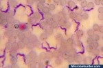
White blood cells obtained from the site of an infection under the microscope.
Microscope info | Citizen Science | Amateur Microscopy

Here I present some photomicrographic images that I made. All pictures are copyrighted and may not be used.
White blood cells obtained from the site of an infection under the microscope.

Here I show you how to observe the pappus of the dandelion. This is the part that carries away the seed with the wind.
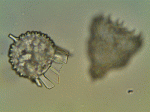
Here we have a focus animation of two radiolaria. The shells of these organisms are made of silica, which is similar to glass.
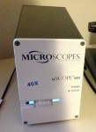
The uSCOPE MXII microscope is a slide digitizer, which allows you to take overlapping pictures to be stitched together into a larger final image.
Automatic white balance can greatly increase the information content of the images, it can also introduce color artifacts, however.
Here is an embedded Youtube video which shows some high speed photography.
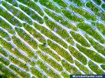
Adding salt water to red onion cells will cause the cytoplasm to lose water by osmosis. The cell’s content shrinks.

Red blood cells do not appear red, when viewed under the microscope.
This parasite is spread over the tsetse fly and causes the sleeping sickness.
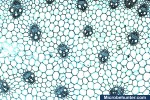
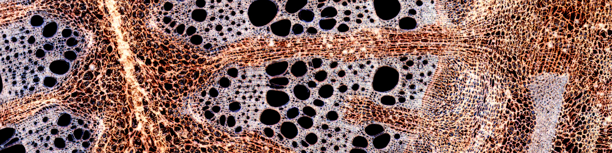
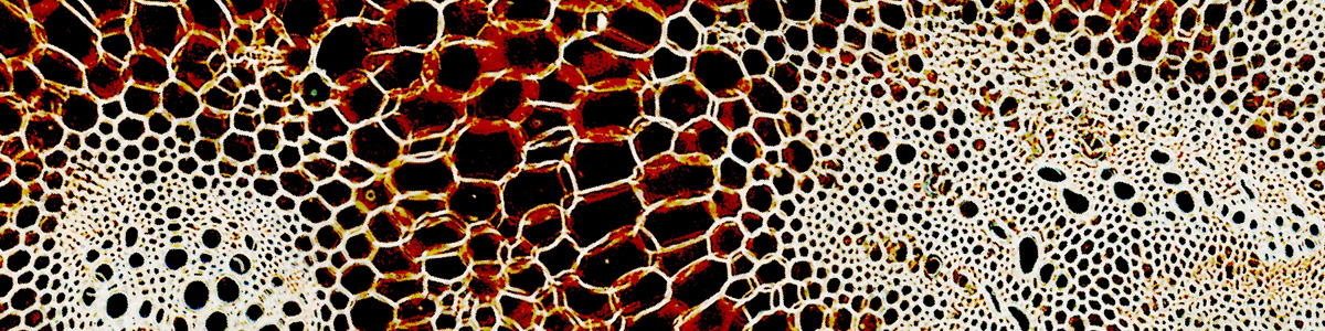
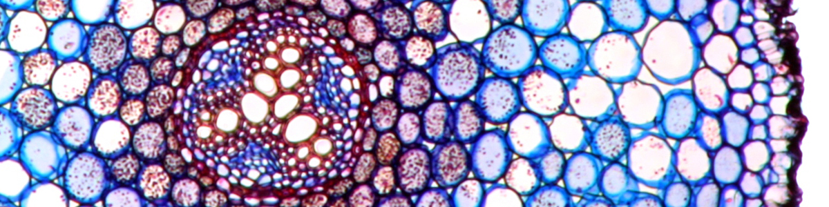
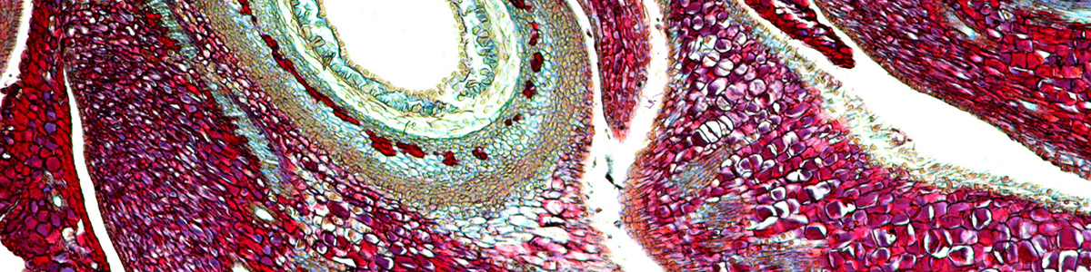
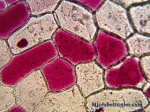
Monocots and dicots can be distinguished by the arrangement of the vascular bundles.
This site uses cookies. By continuing to use the site, you agree to the use of cookies. For more information <a href= more information
The cookie settings on this website are set to "allow cookies" to give you the best browsing experience possible. If you continue to use this website without changing your cookie settings or you click "Accept" below then you are consenting to this.