
Smartphone lens
Thomas Larson from the University of Washington developed a kit which allows you to convert a smart phone into a 150x microscope. He is currently seeking funding to further develop this project. The lens is placed on top of the smart phone camera. The lens adheres to the smart phone camera. It is possible to focus by pressing the slide against the rubber spacers which surround the lens.
Read the original article here
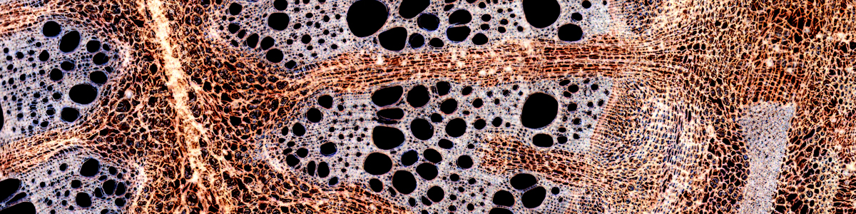
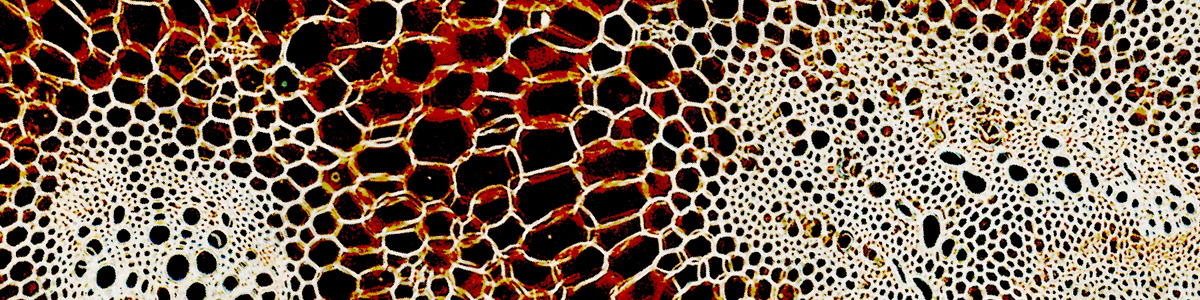
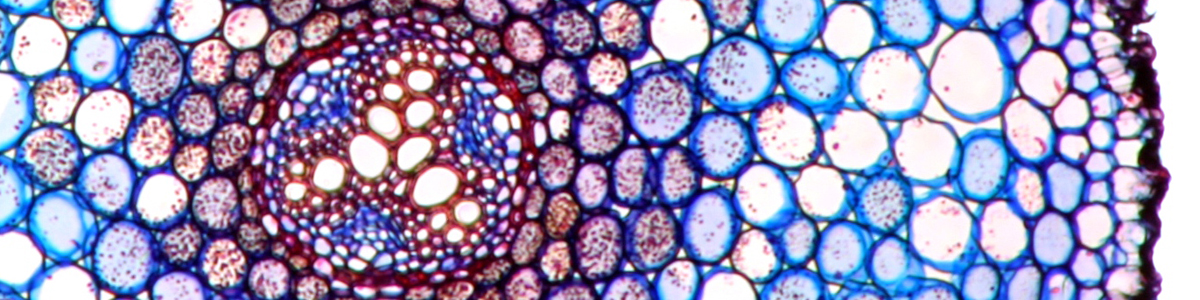
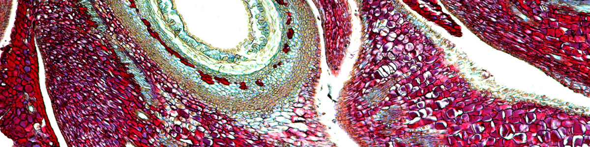
Microphotographs
What is the difference between a photomicrograph and a microphotograph? A photomicrograph is a picture taken through a microscope. Microphotographs are miniature pictures which can be viewed under the microscope. Microphotographs were popular in the 19th century. Miniature photographs of various objects were made on photographic film and then mounted on microscope slides. For viewing these pictures, you had to use a microscope.
Read the original article here
Origami Microscope
Manu Prakash from Stanford University, developed a foldable paper microscope of very low cost. These microscopes can be used to diagnose Malaria in areas where expensive microscopic equipment is not feasible. The “Foldscope” is a single lens microscope, with built-in LED illumination. It is possible to move the slide in two dimensions, much like a mechanical stage. The image can be focused by bending the microscope, which changes the lens to slide distance.
Read the original article here
Video: History of Microscopy
The contributions of Robert Hooke (coined the term “cell”), Anthony van Leuvenhoek and Frits Zernike (invented phase contrast)are well known in the microscopic community. The contribution of Richard Zsigmondy is much less known. The ultra microscope is able to detect particles which are below the resolution limit of the microscope.
Read the original article here
Golub Collection of microscopes
Here is a website from the University of Berkeley with a nice collection of antique microscopes and their description. The website is certainly worth a visit.
Read the original article here
Competition
The winners of an annual micrograph competition will have their pictures displayed on Times Square, New York, between April 25 and 27. The images are impressive both from a visual and from a scientific viewpoint.
Read the original article here
Fast diagnosis
Here we have yet another application that could use the camera of a smart phone as a microscope. Researchers developed a technique for quickly diagnosing the blood of patients for the presence of various bacteria and viruses. A microscope slide is coated with a thin layer of gold which contains small holes. Each one of these “wells” contains antibodies against different bacteria and viruses. If the blood sample contains a corresponding pathogen, then this pathogen will bind against the antibody and a color reaction will be triggered. A smart phone camera could then be used to detect these color changes in the wells. Low cost smart phones can be used to do the analysis, significantly reducing the cost.
Read the original article here
Snowtime
This time-lapse video captures the beauty of a forming snowflake. There is not much more to say. Watch the video, it is worth it!
Read the original article here
Open Textbook
For people who are interested in microbiology, Boundless has published an online textbook under the Creative Commons license. The website also contains introductory chapters on microscopy.
Read the original article here