
Some beginning microscopists are a little disappointed when they look at a sample and are not able to see anything at all, or at least nothing recognizable. There are not many possible reasons for this and you should check each one of the possibilities.
Image not in focus
Quite often the picture is simply not in focus. Maybe you turned the focus knob too far into one diction and you can not see a clear picture anymore. Some people turn the focus knob back and forth and have a problem finding a clear picture. This is a particular problem for the higher magnifications, where the focus can be lost much more easily. If this happens, then you are much faster to simply start over again. Rotate the low magnification objective into place again and re-focus and re-center the image before moving to the higher magnifications.
Visit the Microscopy Shop!
>>> USA Shop | Germany Shop | UK Shop | Canada Shop <<<
As an Amazon Affiliate, I earn a commission but it does not cost you more.
Wrong focus plane
Maybe you are able to see a sharp image, but not of the object that you want to look at. In this case you probably focused not on the specimen itself, but on the dust on the surface of the slide or cover glass. In this case it is best to start over and re-focus with the low magnification objective in place. It may also be the case that you focused on the dust which is located on the lens of the condenser, which is beneath the slide. Simply move the slide back and forth. If the dust stays stationary and does not move, then you did not focus on the slide.
Slide not centered
This is another common problem. Are you actually looking at the specimen or are you trying to observe a part of the slide which does not contain the object of interest? Maybe you can not see anything because you are looking at a place on the slide which does not contain a specimen at all. I have even seen extreme cases, of people looking at the paper label of the slide instead of the specimen! Always center the slide before starting the observation. Look at the slide on the stage with the lamp turned on. You should be able to see the specimen in the light right above the condenser.
Objective not locked in place
This seems to be a trivial reason, but even this can cause problems. Always rotate the objective fully into place, until you hear the “click” sound. This is a particular problem with stereo microscopes where you can not see the position of the objectives very well.
The sample is of too low concentration
This means that you are only observing a clear liquid without many cells or other particles. As a general rule of thumb, if you are able to see through the sample without any problems, then you will also not be able to see anything under the microscope. Clear pond water is a typical case. Most living things do not float around in the water but are attached to rocks, decaying leaves or plants in the pond.
Specimen too dark
if you place a large and dark specimen on the stage, then the light of the microscope is not able to pass though the object. You will not be able to see anything except a dark shadow without much detail. In this case you must either cut the specimen into thin sections, tear it apart or squash it. If you can not do these preparation techniques, then you have to use a stereo microscope. Rock and mineral samples are such a typical case.
Condenser iris diaphragm setting not correct
A diaphragm which is too open will result in an image which has low contrast and a low depth of field. Some specimens may be difficult to see under these conditions. Close the diaphragm. You do this by moving it into the position which gives you the darkest image. You can then turn up the light.
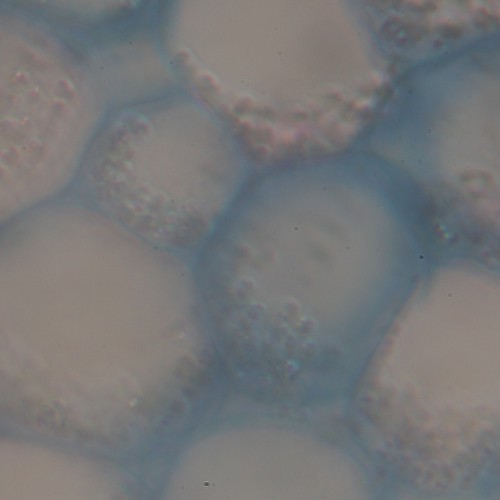
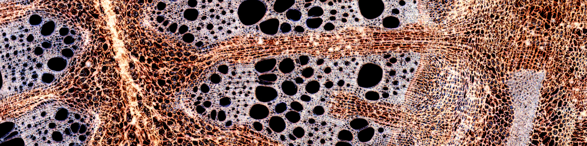
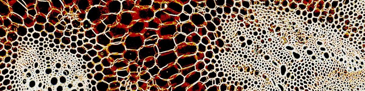
Objective dirty
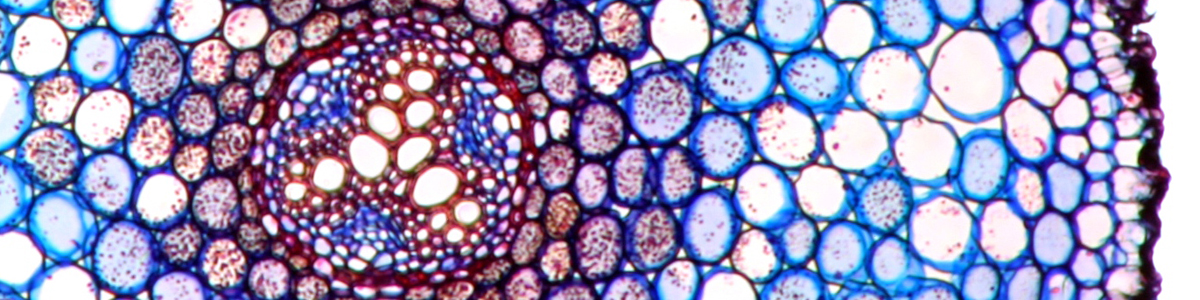
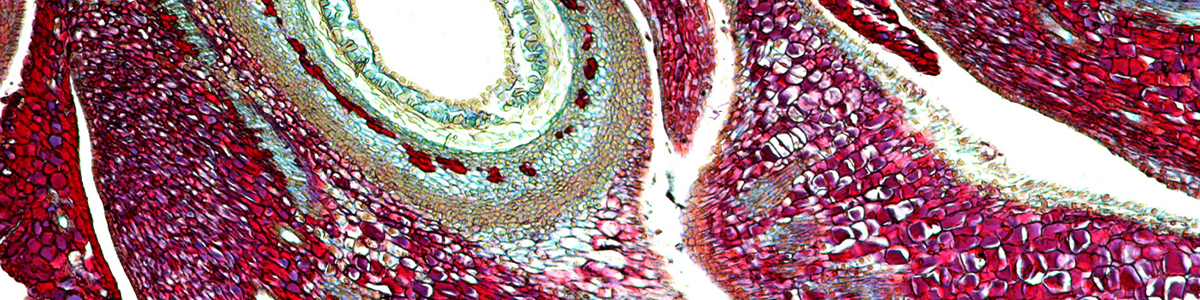
A dirty objective can greatly reduce the image quality. This can be the case, when a non-oil immersion objective becomes covered with immersion oil and if it is not cleaned. Dust and dirt will stick to the oil and block the light. The two pictures below compare the effect. The two images are both taken with a 40x objective and the objective on top is the one that was dirty. The image lack both contrast and resolution.