
This time I’d like to talk about a topic which is only indirectly related to microscopy: macro imaging.
Microscope info | Citizen Science | Amateur Microscopy

Here I present some photomicrographic images that I made. All pictures are copyrighted and may not be used.
This time I’d like to talk about a topic which is only indirectly related to microscopy: macro imaging.
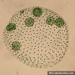
Volvox is a fresh water green algae and a member of the Chlorophyta.

This is one of the first tries taking pictures with my new Sigma objective, and I have to admit that I’m very satisfied with the lens.

Spring time is pollen time! Here are two images of Ranunculus repens (the Creeping Buttercup or Creeping Crowfoot) pollen.

Today I’d like to show you a nice microscopic picture, which I took several years ago of two human hair.

Phase Contrast microscopy makes specimens of low contrast appear with greater contrast.

Trichinella spiralis is the smallest nematode parasite in humans. It causes the disease trichinosis. It is also one of the most wide spread parasites of the world. It can be contracted by eating raw or half-cooked pork or wild game animals.
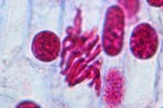
Mitosis stages of the lily flower. The chromosomes are well visible.
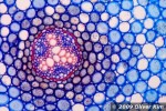
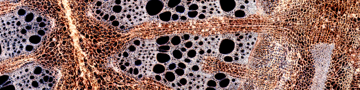
Vascular tissue of a Buttercup, Ranunculus, root.
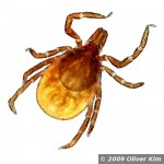
A tick in dark field. Ticks are parasites that feed on blood. They are known to transmit a variety of diseases, such as Lyme disease (borreliosis) and tick-borne encephalitis.
This site uses cookies. By continuing to use the site, you agree to the use of cookies. For more information <a href= more information
The cookie settings on this website are set to "allow cookies" to give you the best browsing experience possible. If you continue to use this website without changing your cookie settings or you click "Accept" below then you are consenting to this.